/Acute%20Otitis%20Media%20-%20Perforation%2C%202.webp)
/Acute%20Otitis%20Media%20-%20Perforation%2C%201.webp)


Description
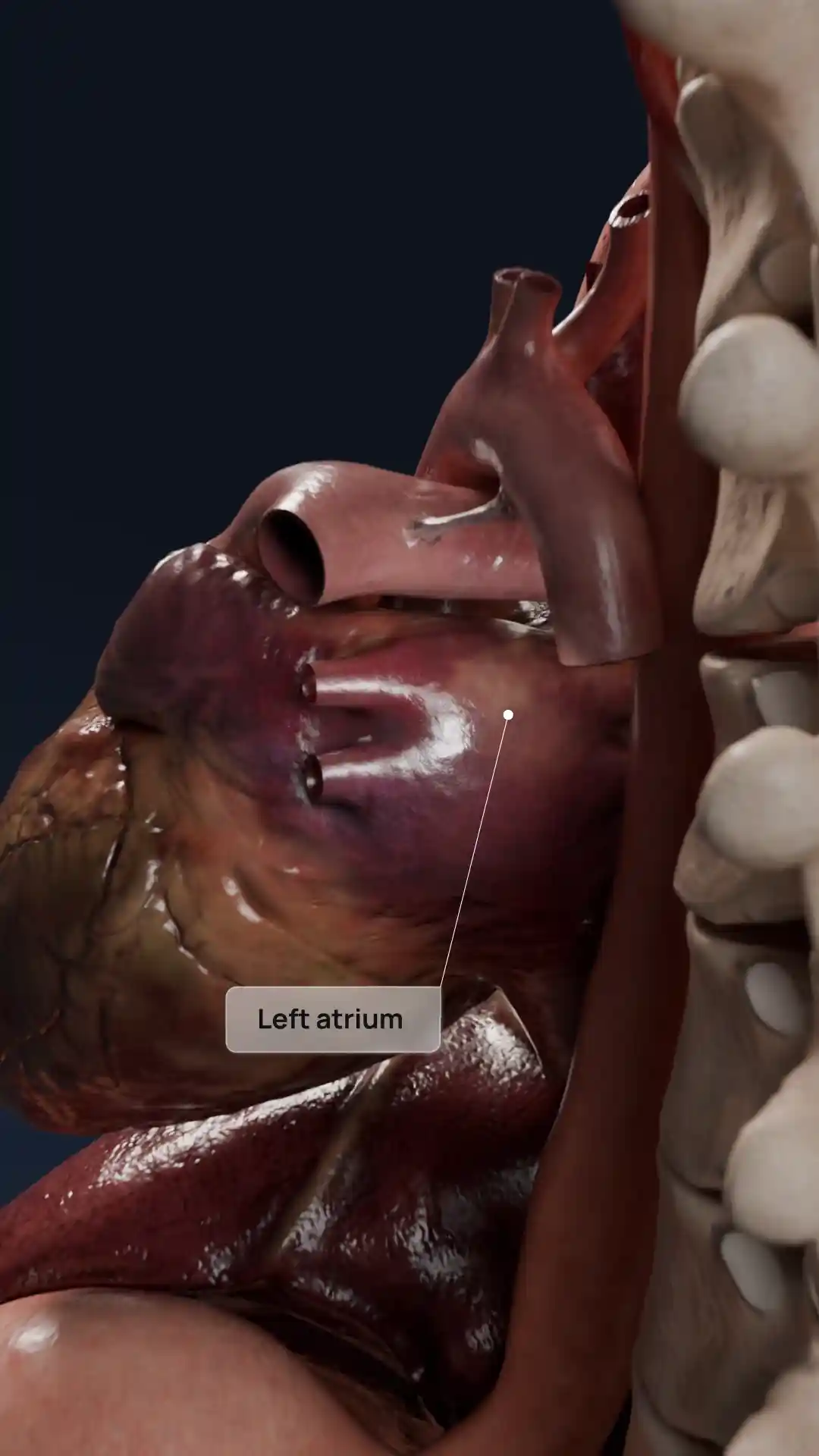
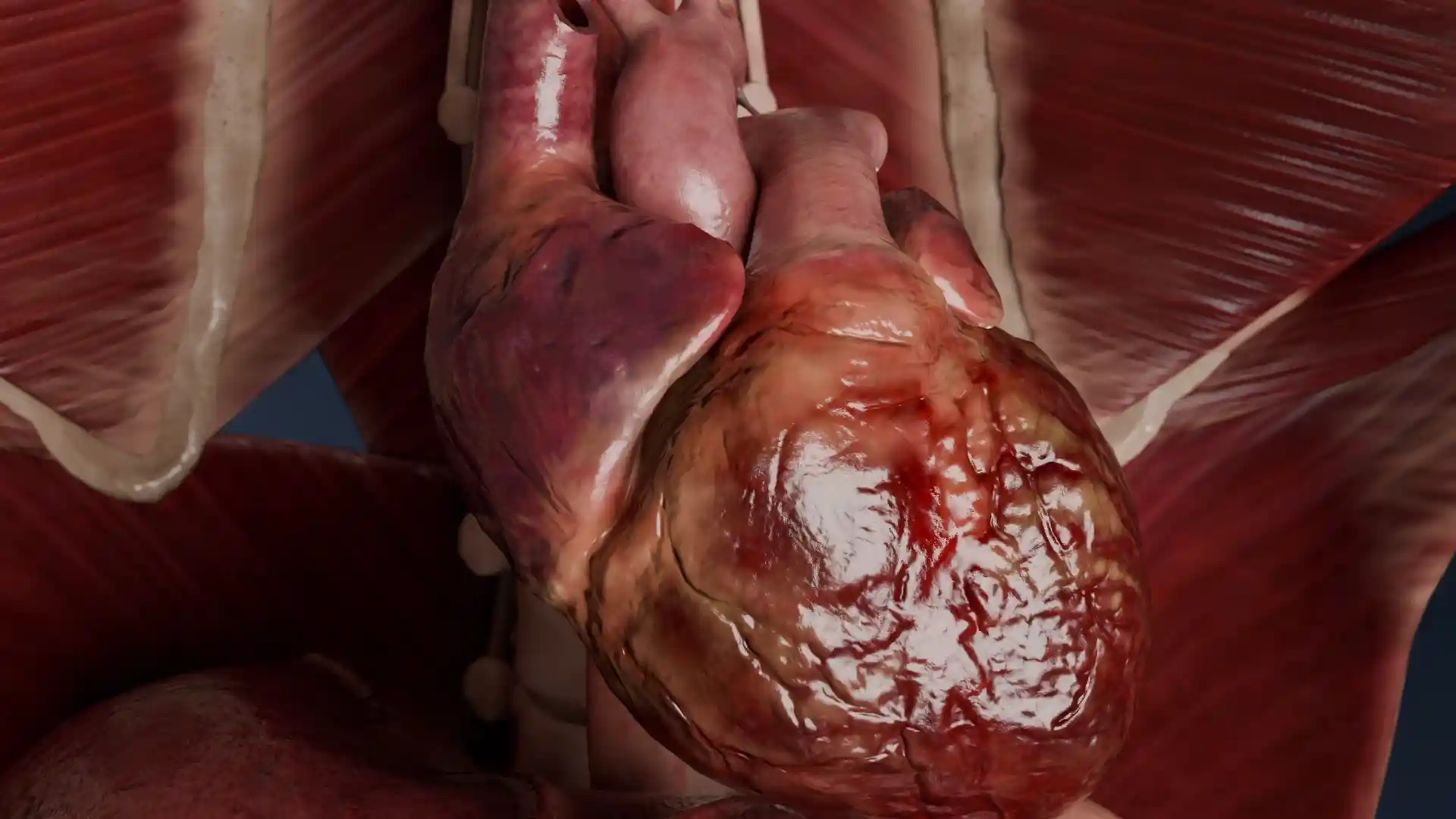
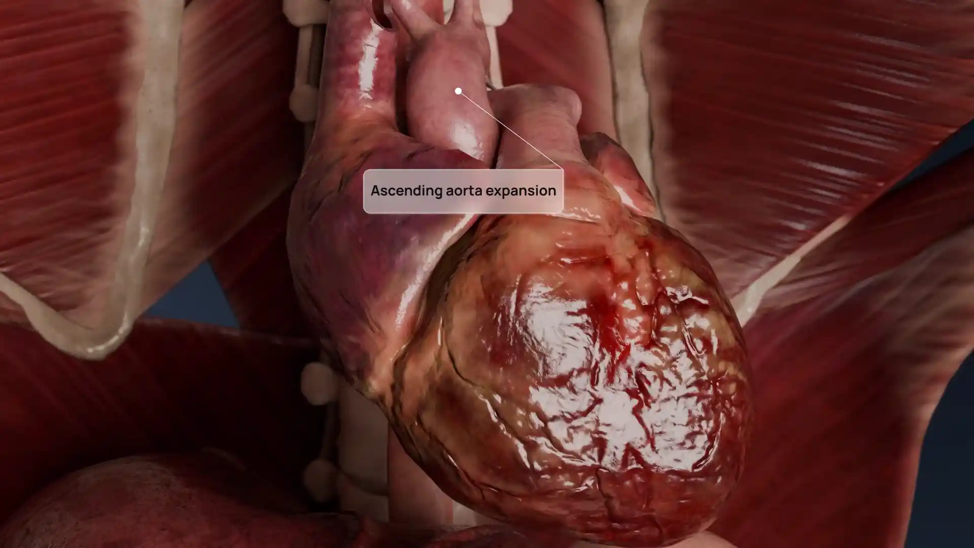
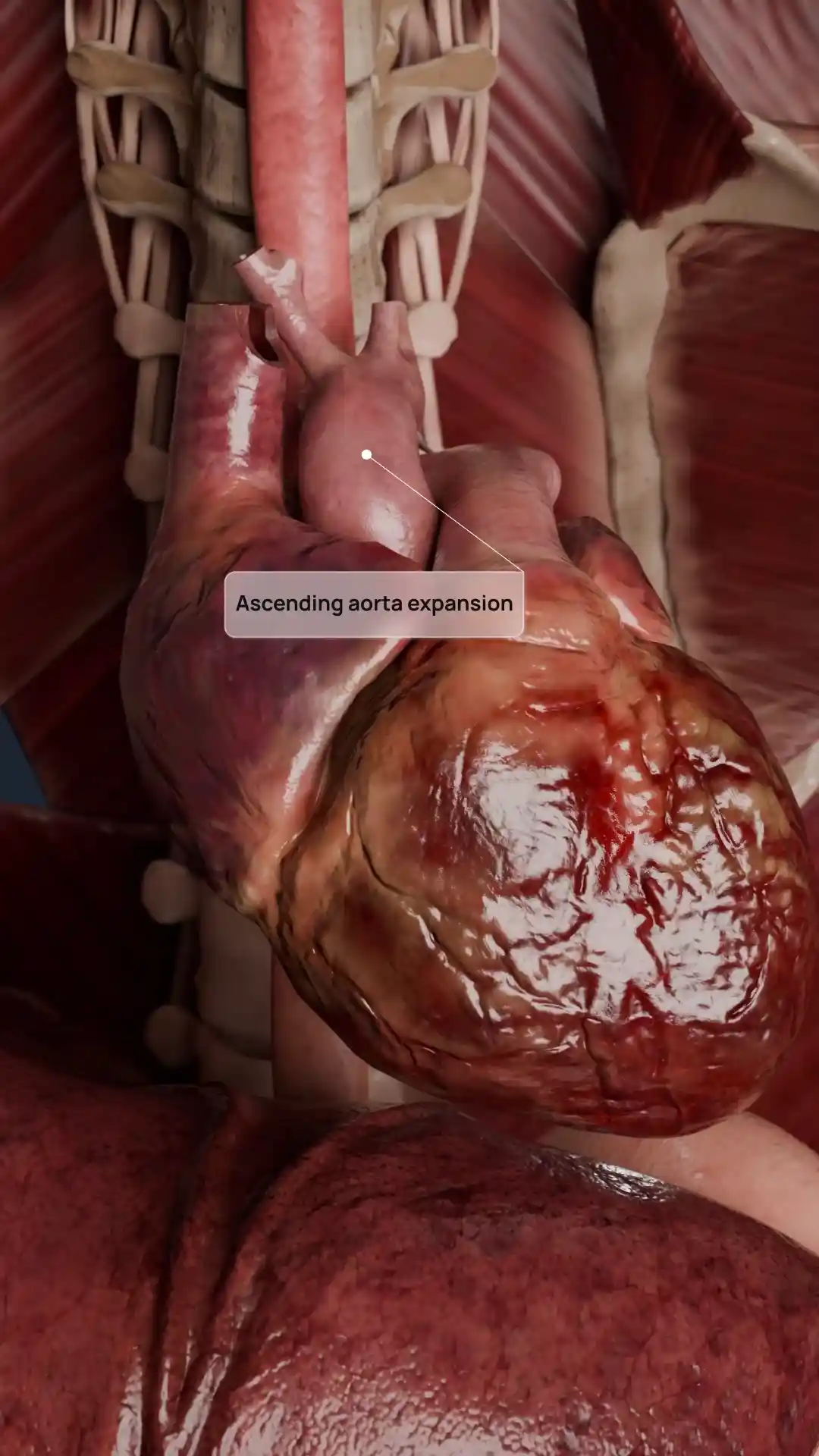
This detailed 3D illustration showcases Type II acquired tricuspid valve disease, characterized by structural distortion and displacement of the valve leaflets. The sagittal heart section allows clear visualization of the thickened, retracted valve tissue and impaired leaflet coaptation. The image highlights key anatomical relationships, including the chordae tendineae, papillary muscles, and right atrioventricular orifice, offering a comprehensive look at both function and pathology. Ideal for cardiology education, clinical case presentations, and patient communication, this visual model provides a high-fidelity reference for understanding the mechanical and morphological changes associated with tricuspid valve dysfunction.
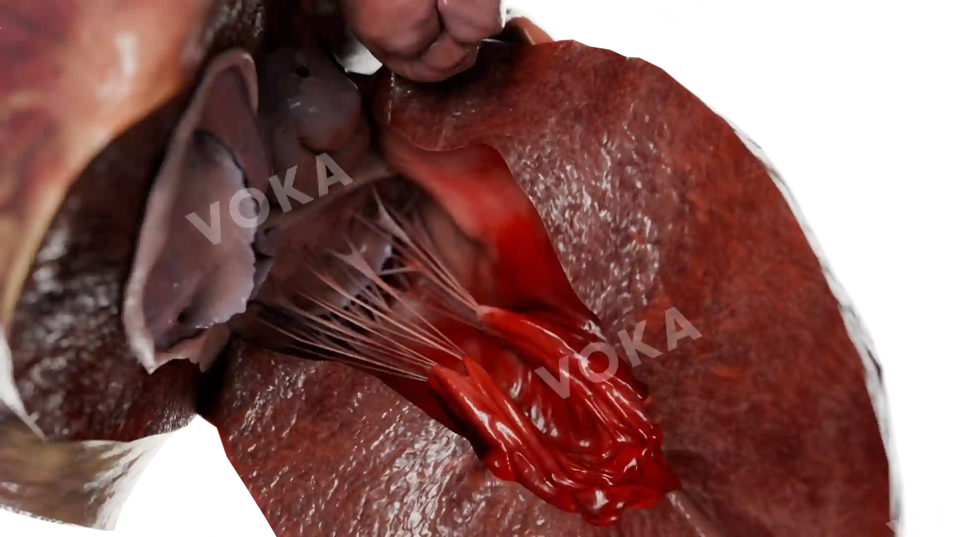
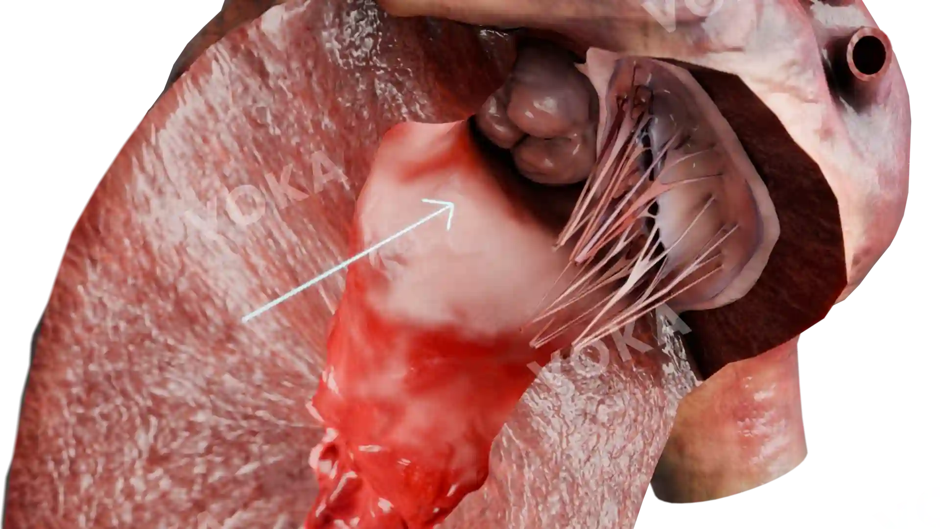
Acquired tricuspid valve diseases type II image - 30092
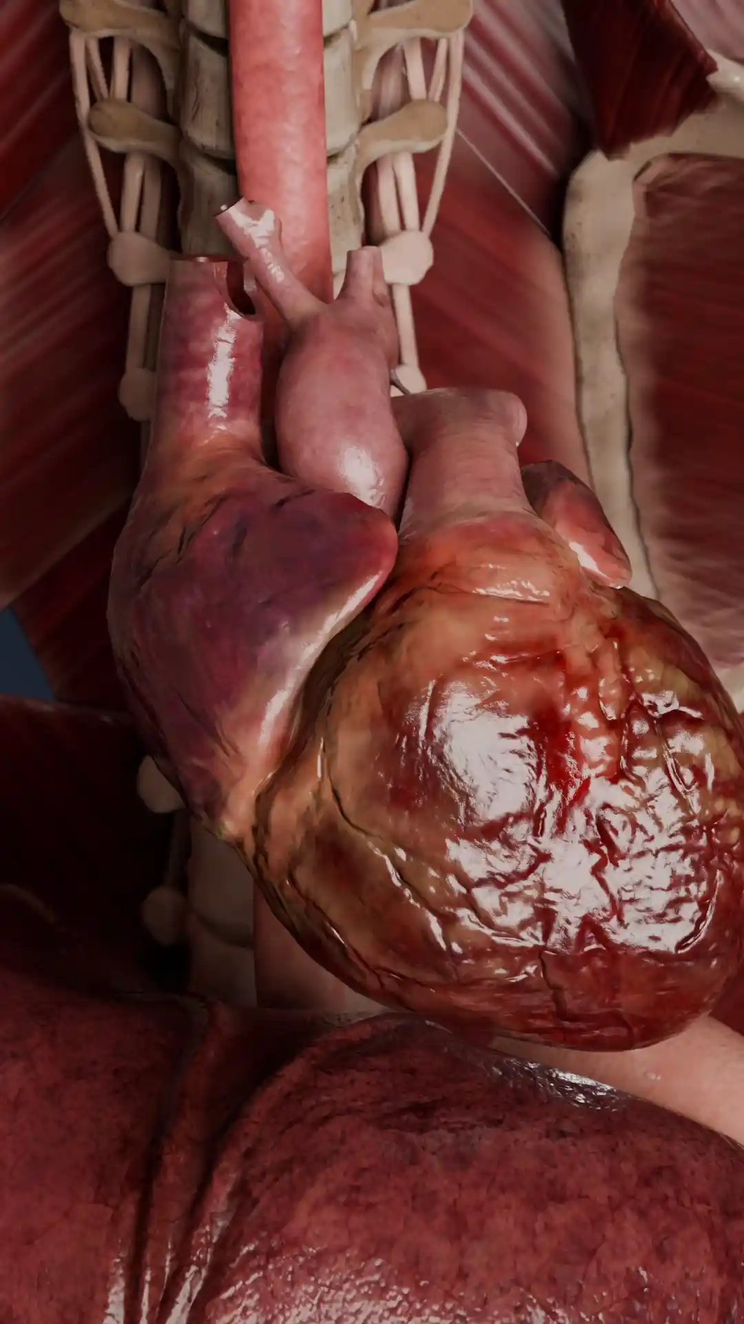
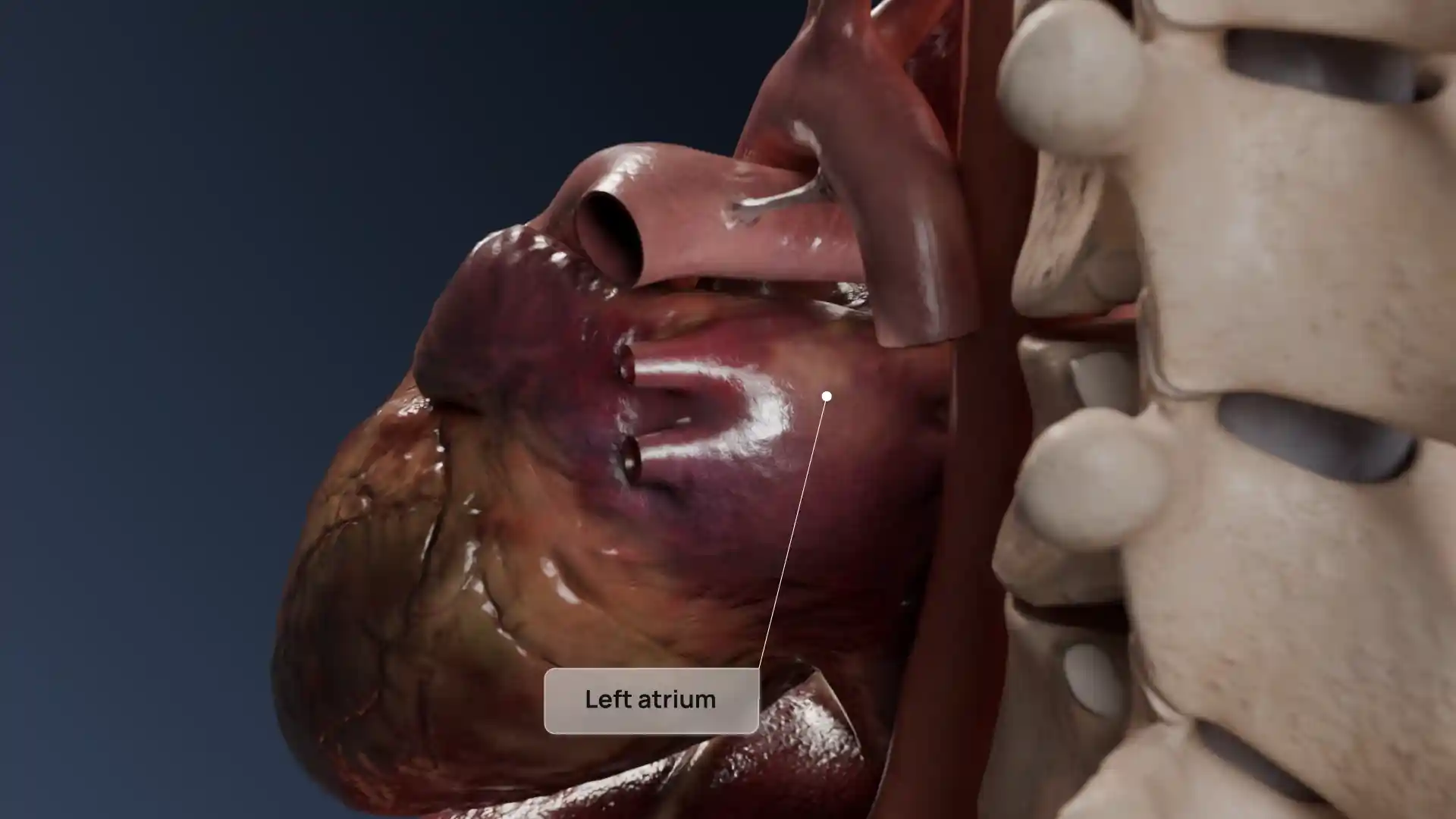
Cardiovascular system
Select license
More information
Details
Item successfully added to the cart