/Acute%20Otitis%20Media%20-%20Perforation%2C%202.webp)
/Acute%20Otitis%20Media%20-%20Perforation%2C%201.webp)


Description
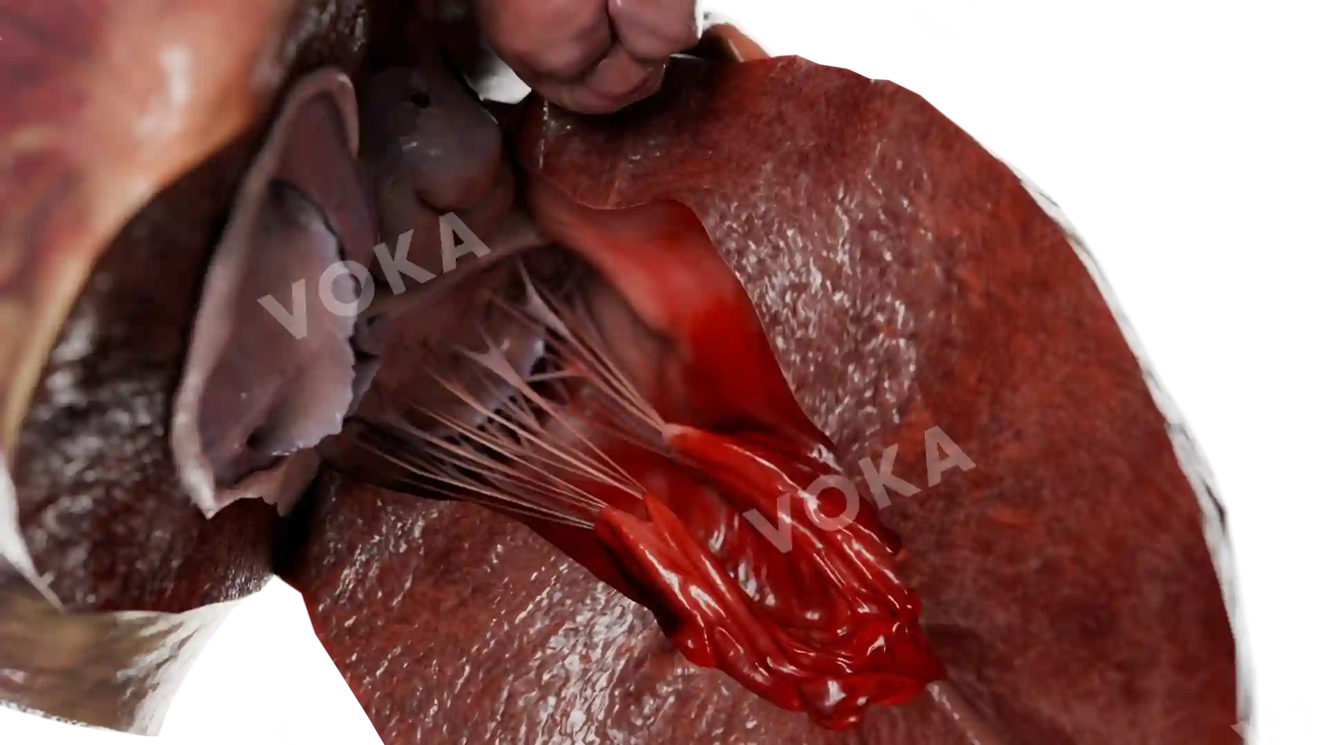
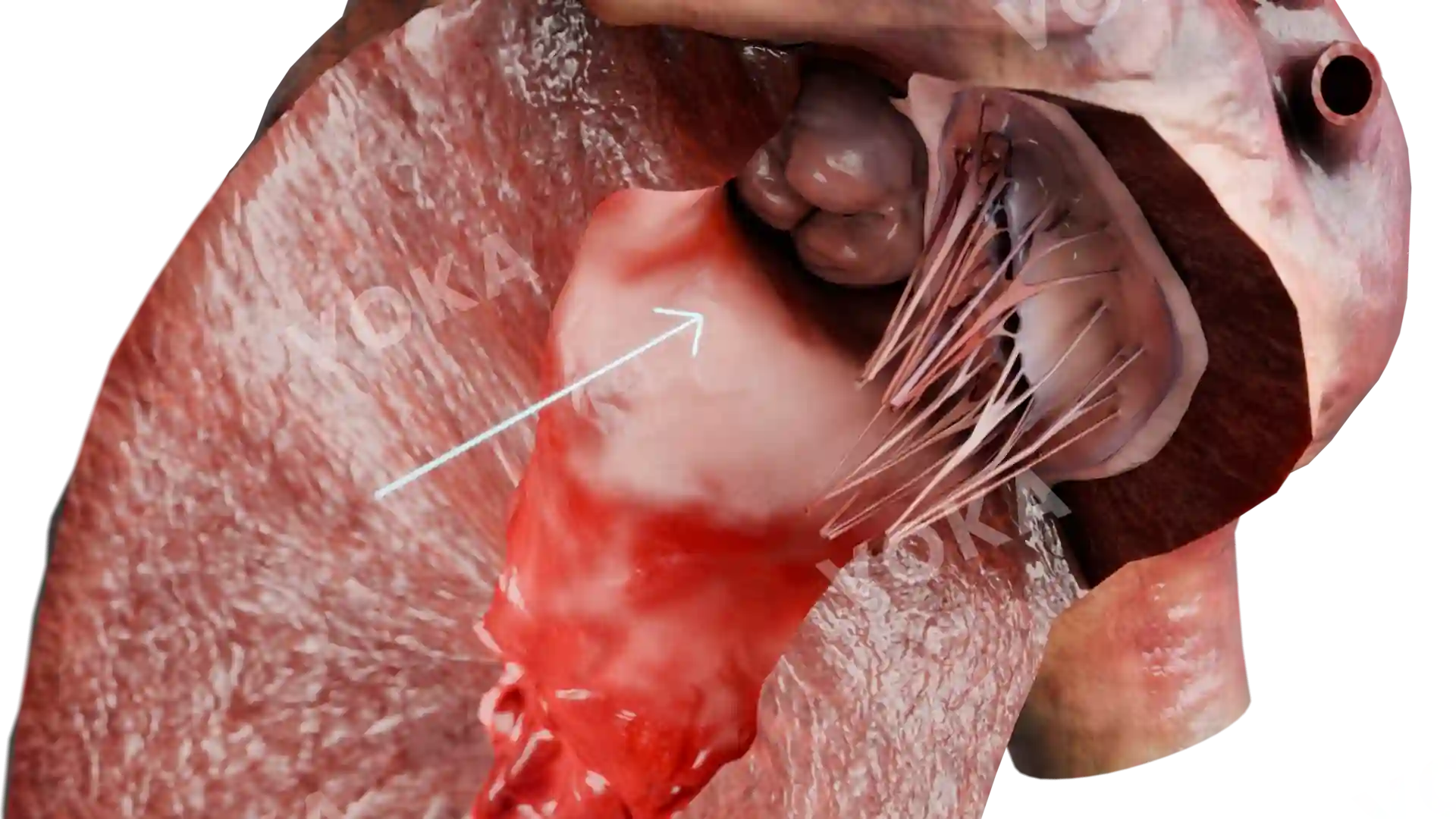
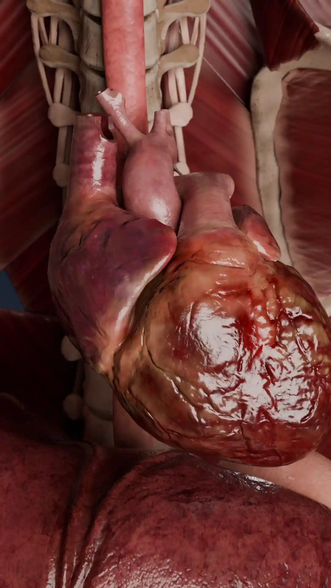
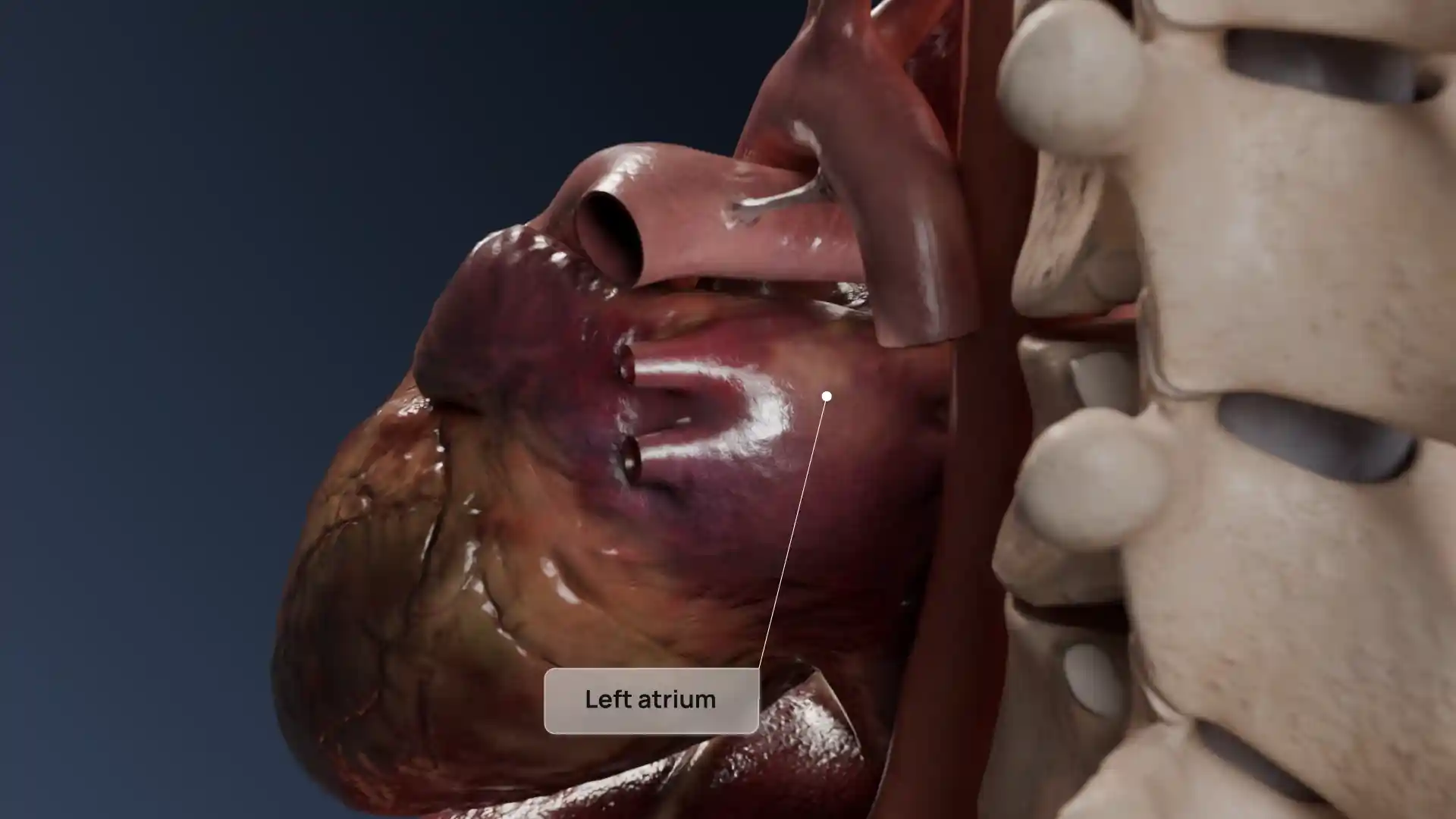
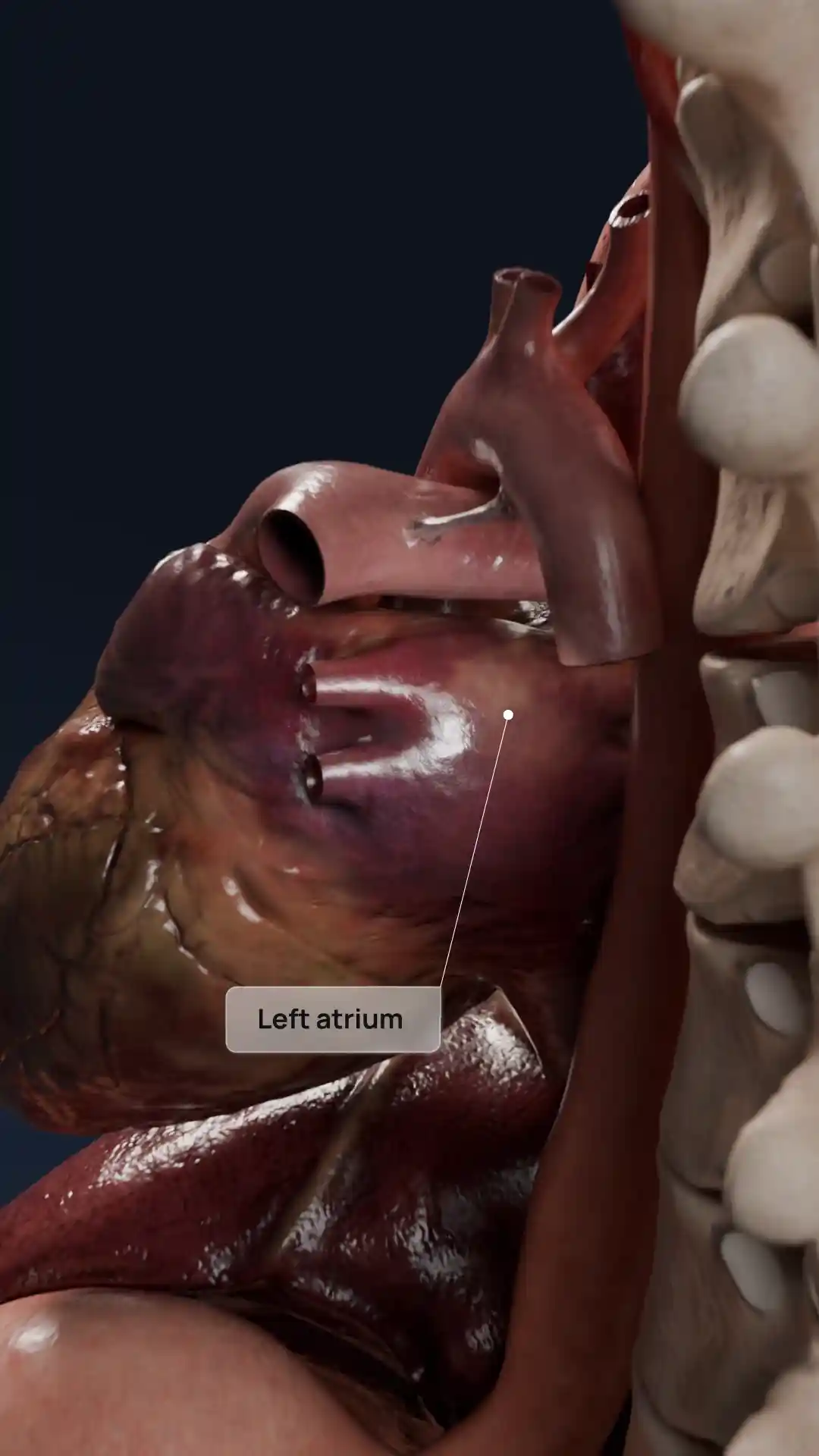
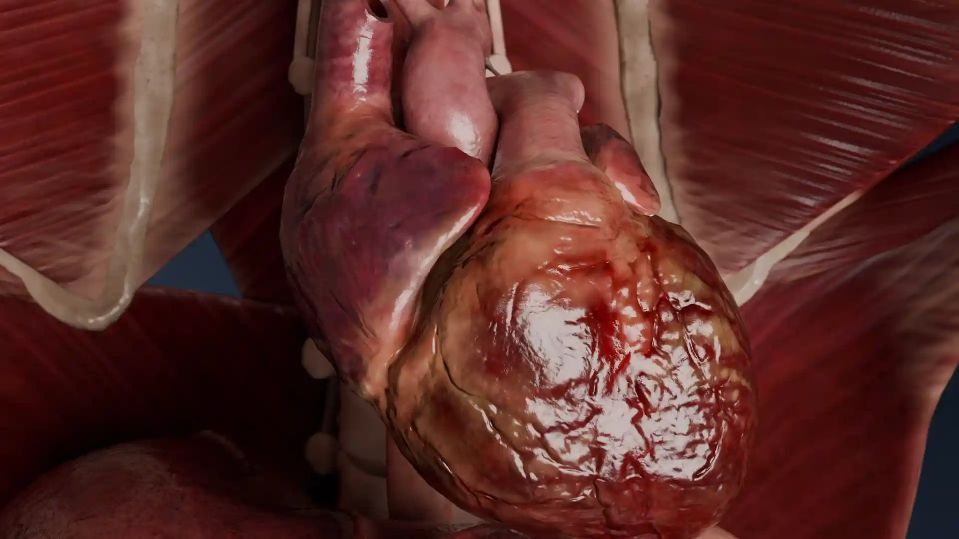
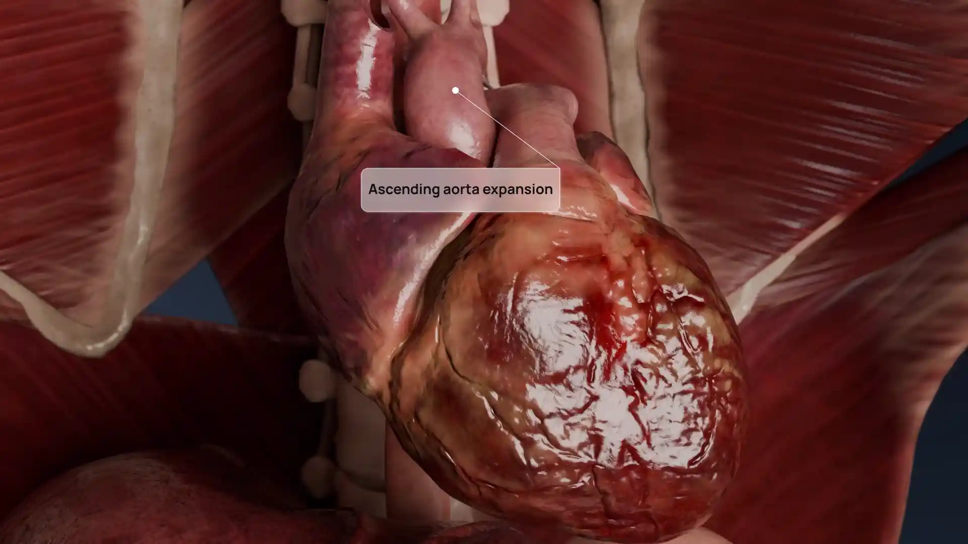
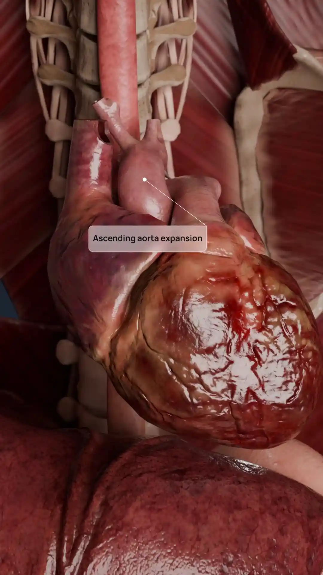
This anatomical visualization depicts Type IIIb acquired tricuspid valve disease, commonly associated with ventricular dilation and leaflet tethering. The 3D sagittal section reveals restricted leaflet motion due to displaced papillary muscles and stretched chordae tendineae, while the valve annulus appears dilated. These structural changes impair proper coaptation, contributing to functional tricuspid regurgitation. Designed with clinical accuracy and based on real imaging data, this model provides a clear and dynamic perspective on the underlying mechanisms of the pathology. It serves as a valuable tool for cardiology education, surgical planning, and patient communication.
Acquired tricuspid valve diseases type IIIb image - 30093
Cardiovascular system
Select license
More information
Details
Item successfully added to the cart