/Acute%20Otitis%20Media%20-%20Perforation%2C%202.webp)
/Acute%20Otitis%20Media%20-%20Perforation%2C%201.webp)


.webp)
Description
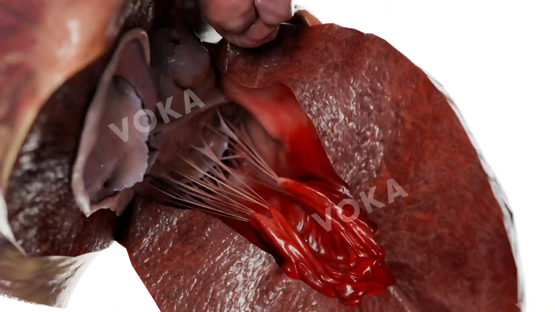
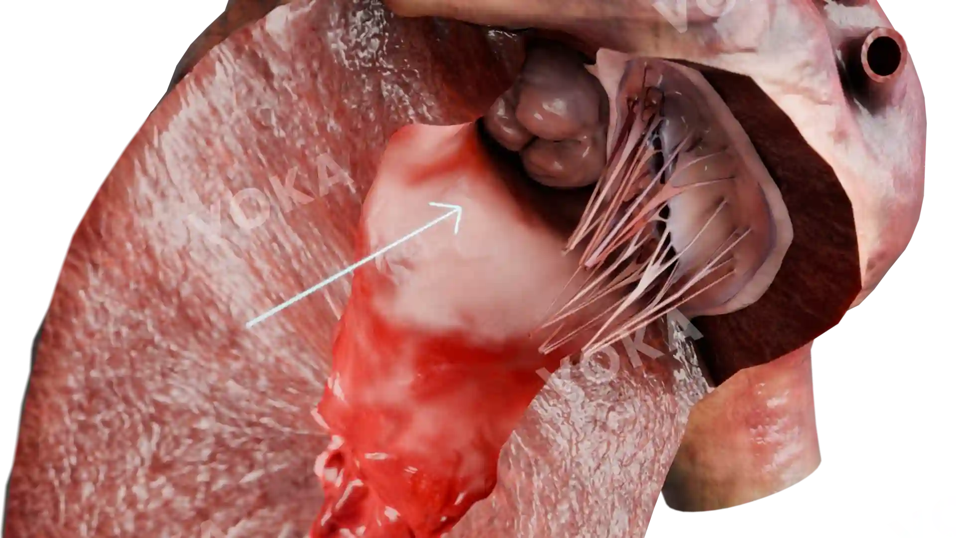
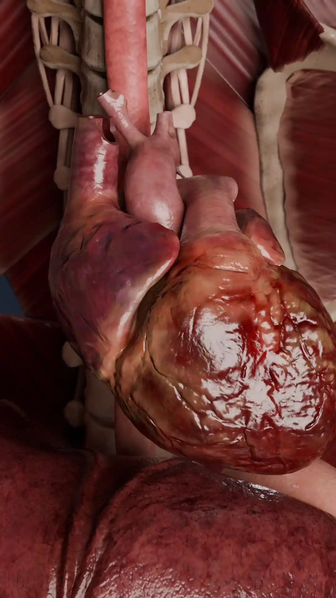
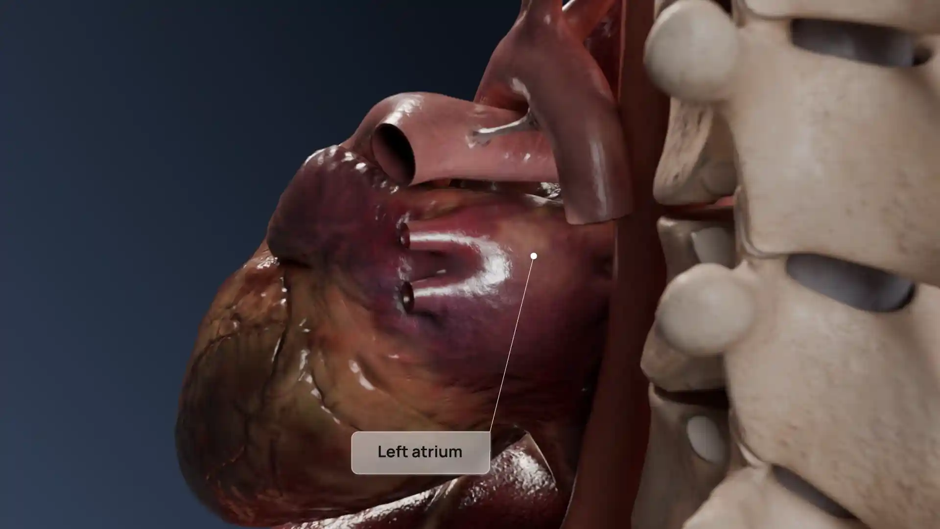
This detailed anatomical visualization illustrates an atrial septal defect (ASD), a congenital heart condition characterized by an abnormal opening in the atrial septum, the wall separating the heart's two upper chambers. The image highlights how this defect allows oxygen-rich blood from the left atrium to mix with oxygen-poor blood from the right atrium, leading to increased pulmonary blood flow and potential strain on the heart over time. This visualization is invaluable for medical education, cardiology research, and clinical reference, offering a clear view of the defect’s role in complications such as pulmonary hypertension, heart rhythm abnormalities, and heart failure. Understanding ASD is crucial for early diagnosis and treatment, whether through surgical correction or catheter-based interventions.
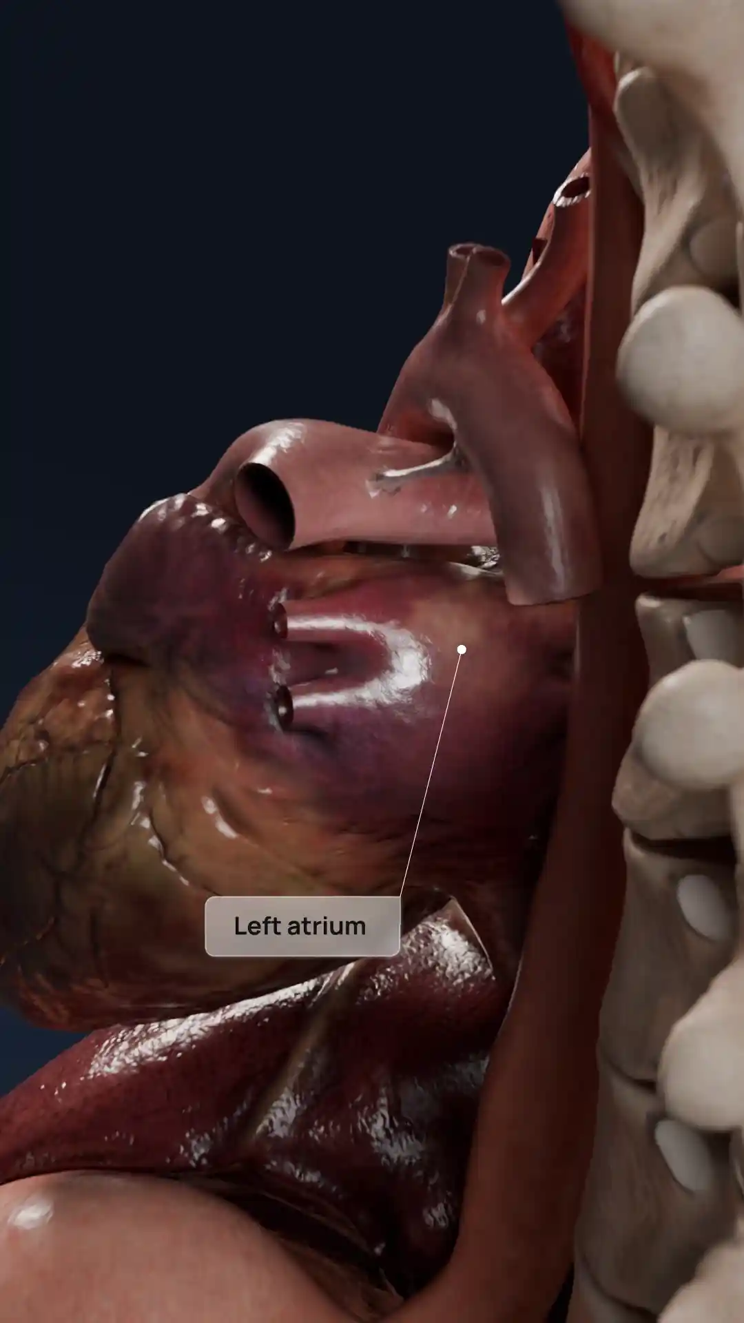
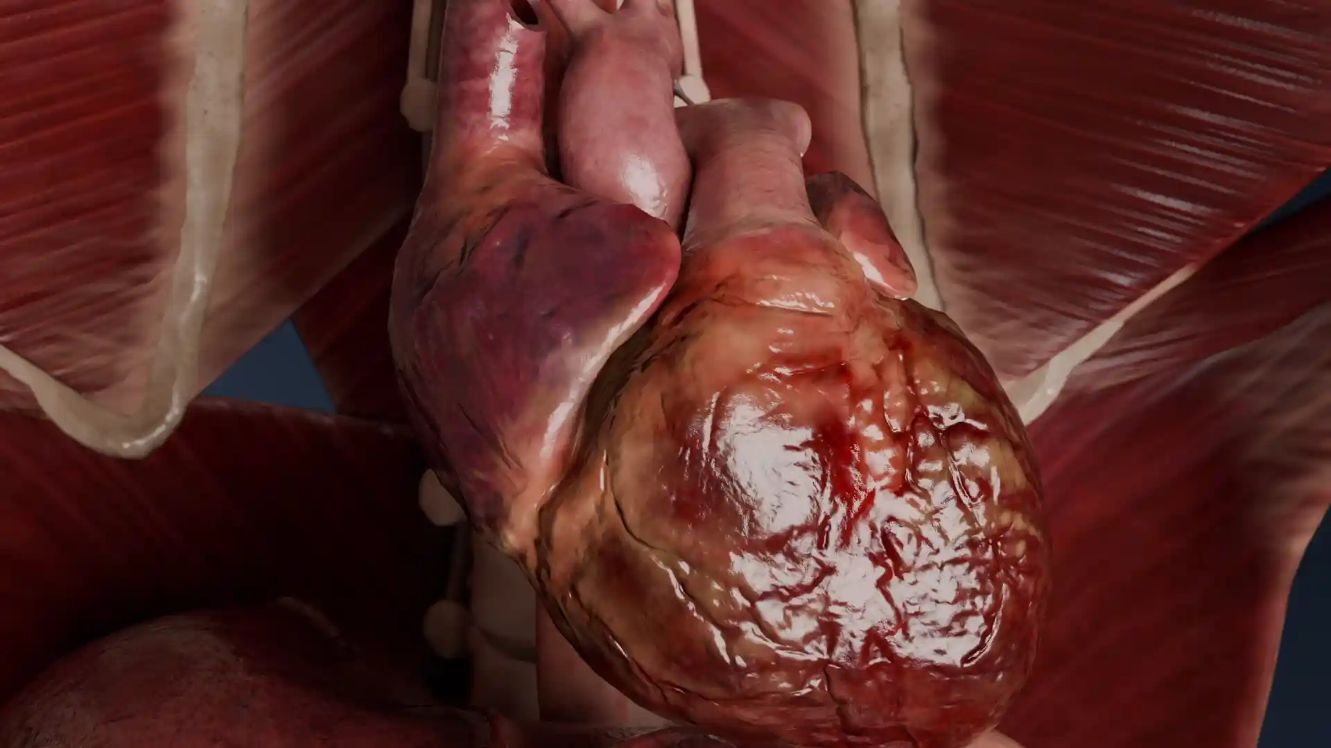
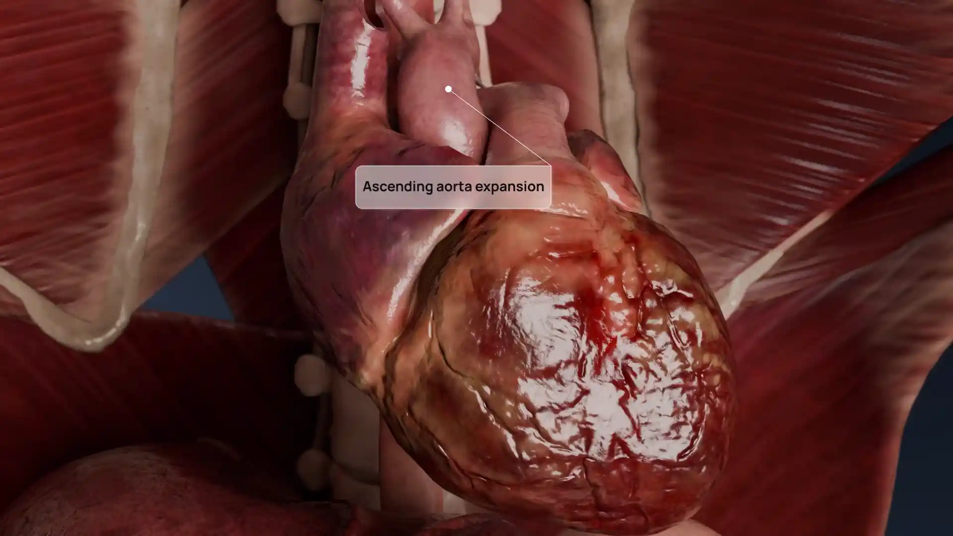
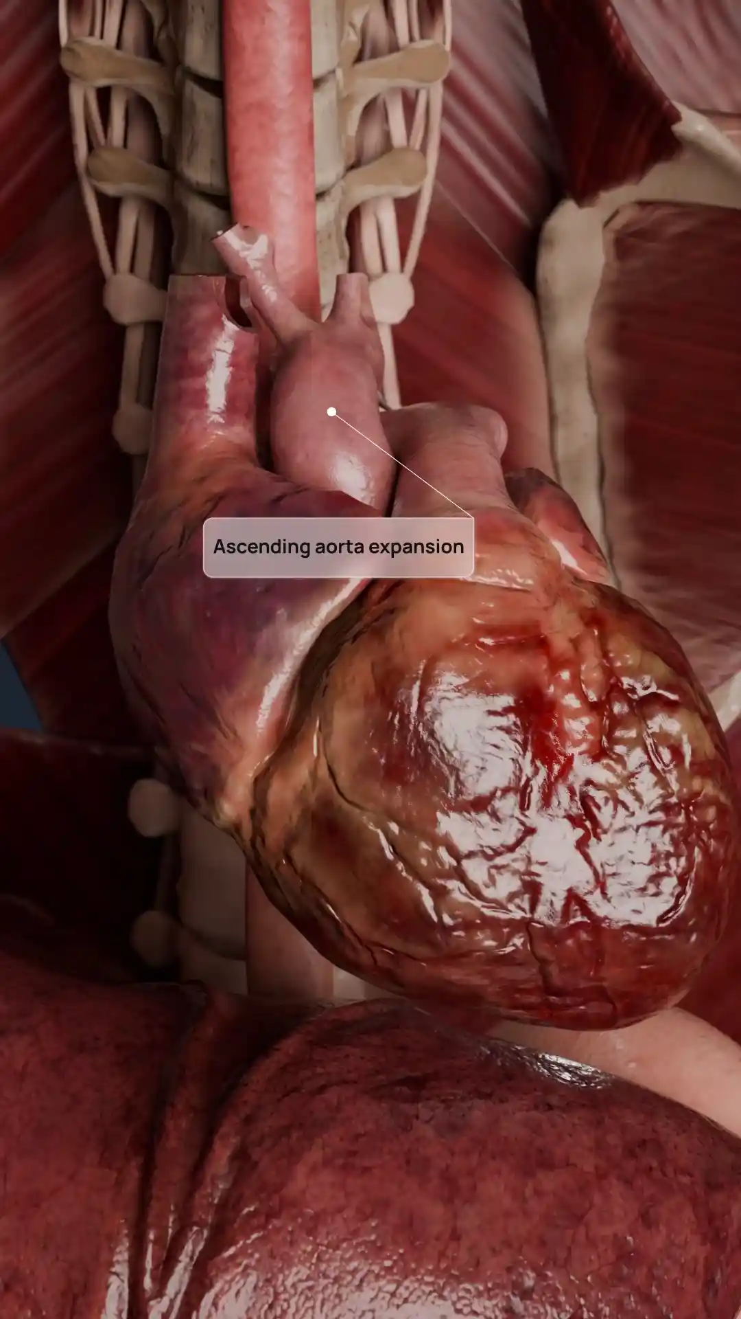
Atrial septal defect (ASD) image - 25012
Cardiovascular system
Select license
More information
Details
Item successfully added to the cart