/Acute%20Otitis%20Media%20-%20Perforation%2C%202.webp)
/Acute%20Otitis%20Media%20-%20Perforation%2C%201.webp)


Description
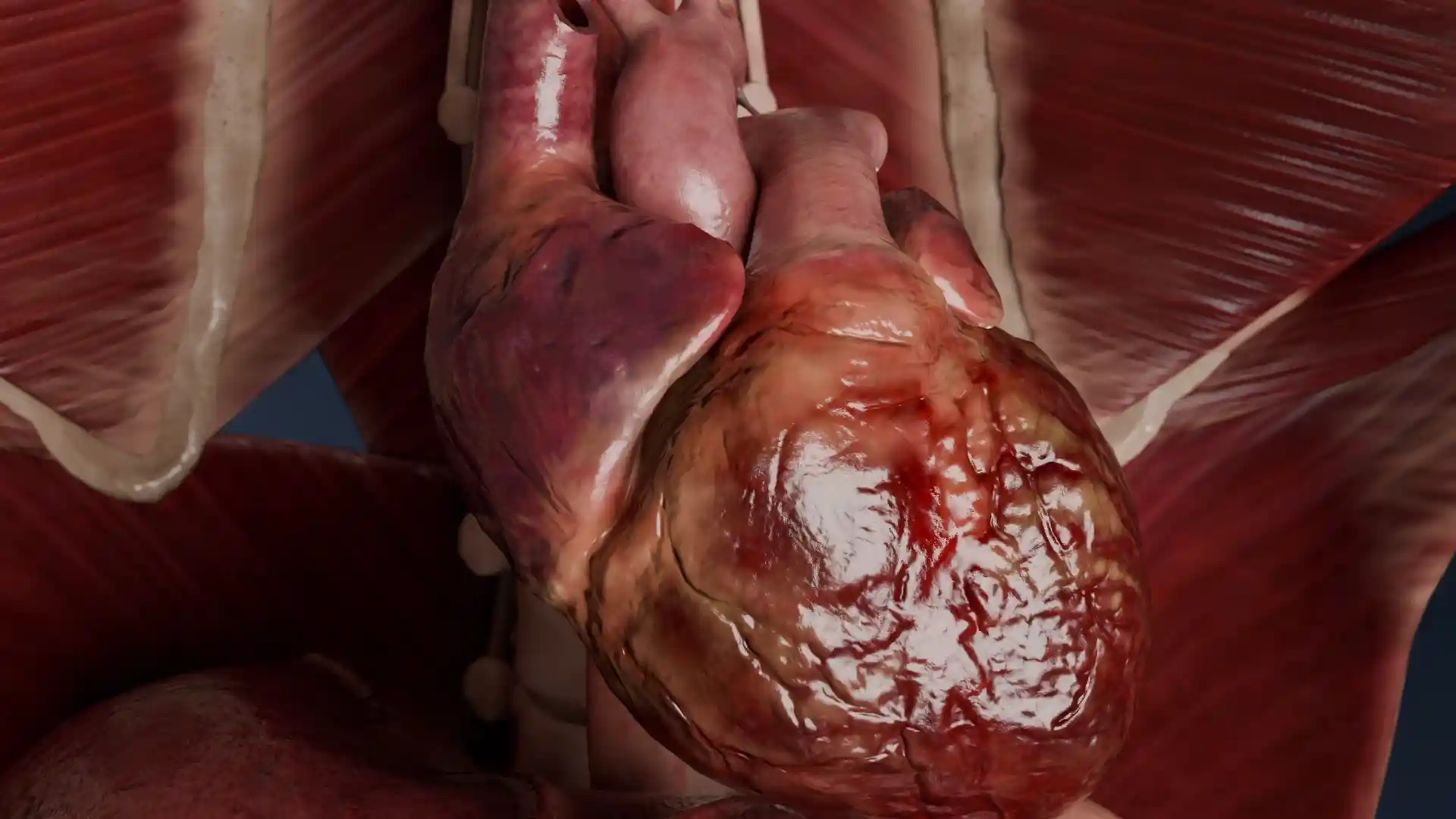
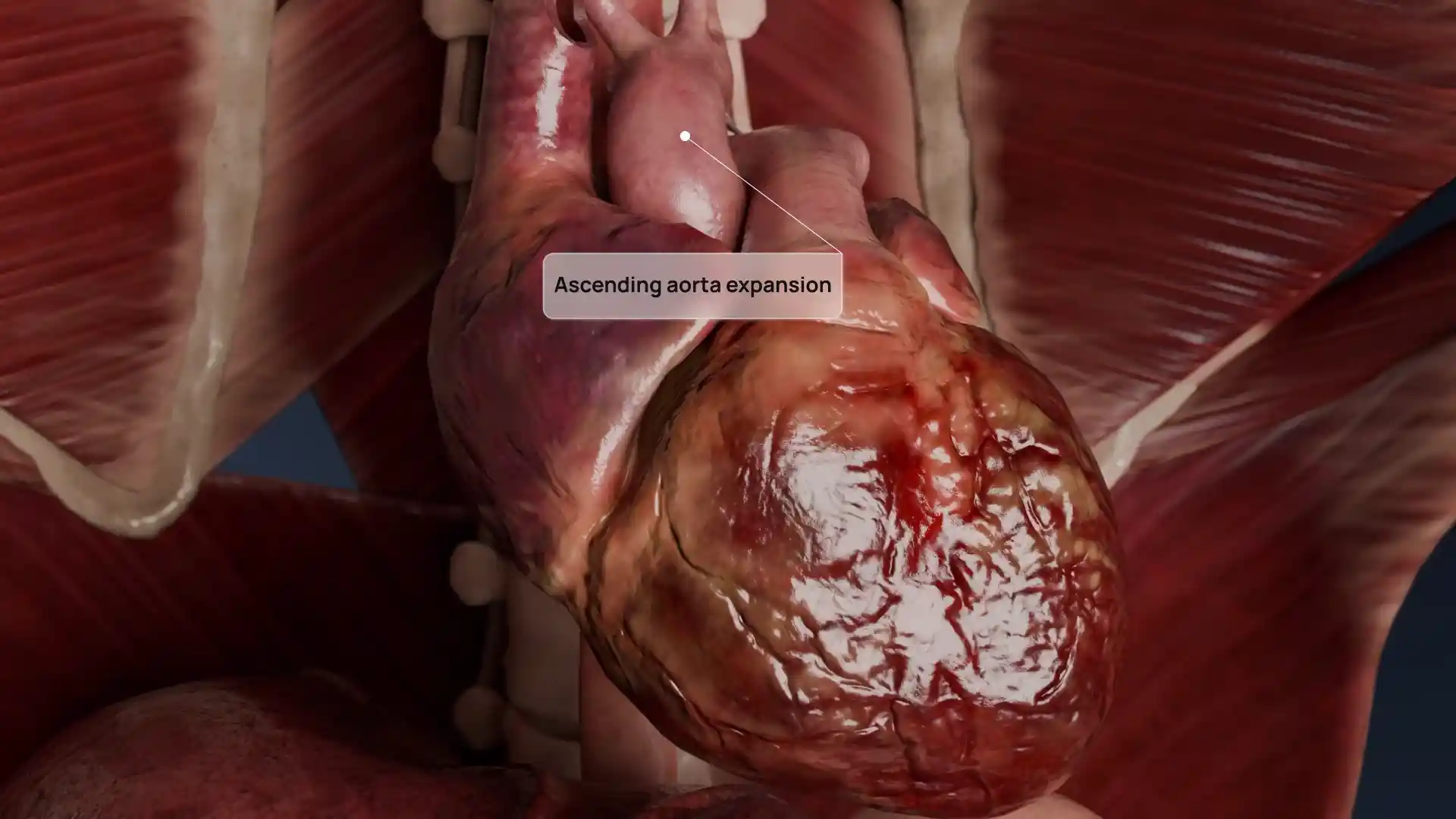
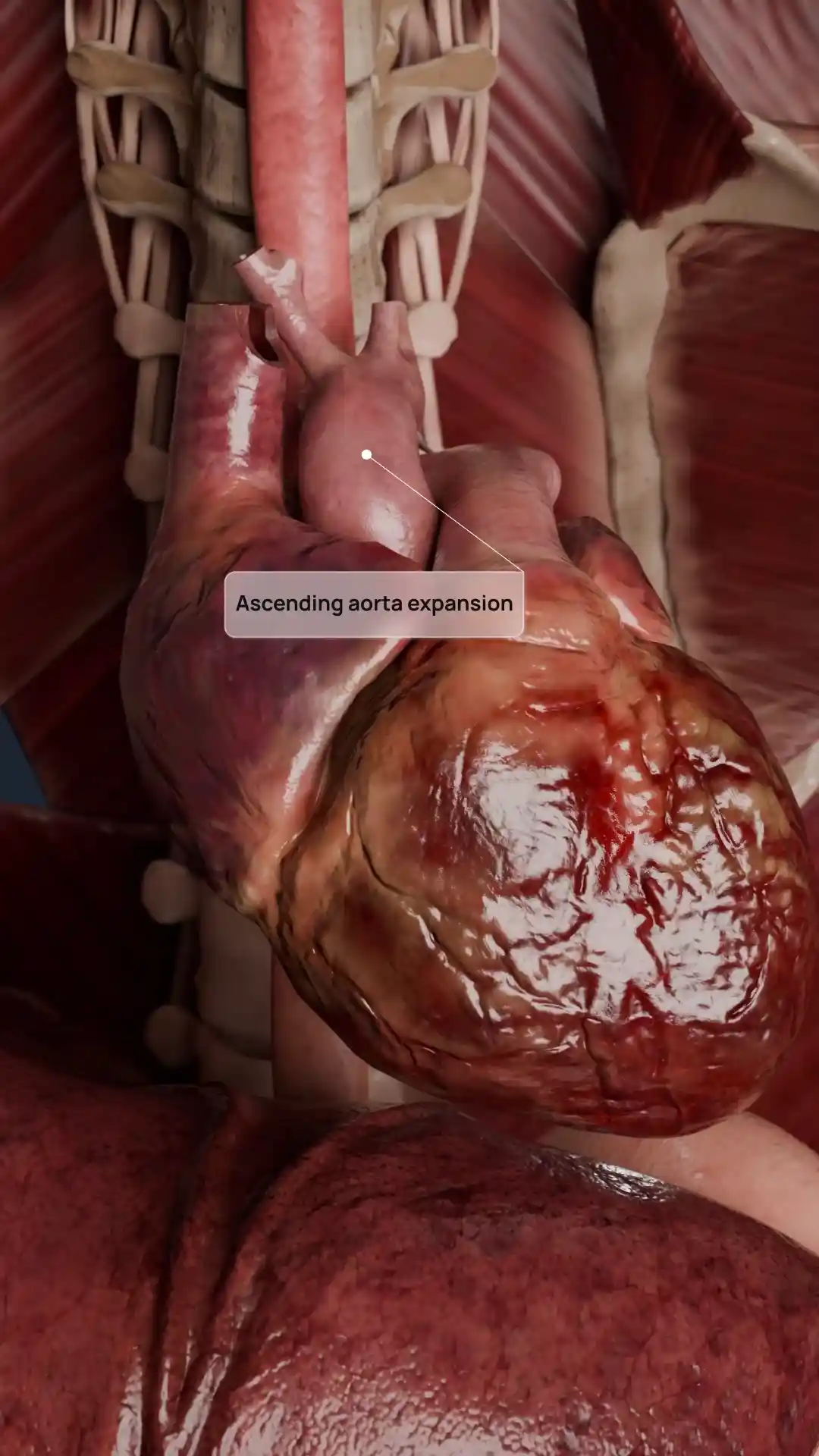
This detailed anatomical rendering captures a close-up view of a classic vascular pathology: a dissection flap of the intima located just distal to the origin of the left subclavian artery. The image vividly highlights the separation between the true and false lumens of the aortic wall, a hallmark of aortic dissection. The clean, cross-sectional visualization offers a rare glimpse into the internal mechanics of this life-threatening condition, where blood penetrates between the layers of the arterial wall, forming a false channel. Whether you're teaching advanced cardiovascular pathology or building a presentation on aortic emergencies, this illustration is a valuable asset for demonstrating one of the most critical vascular conditions in modern medicine.
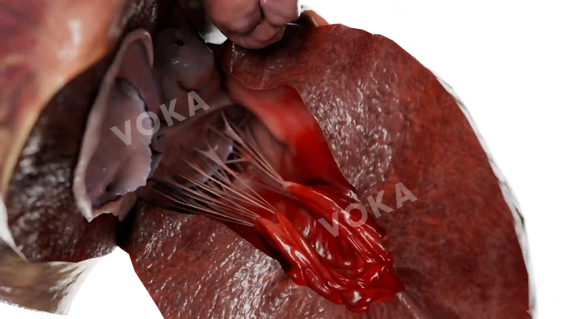
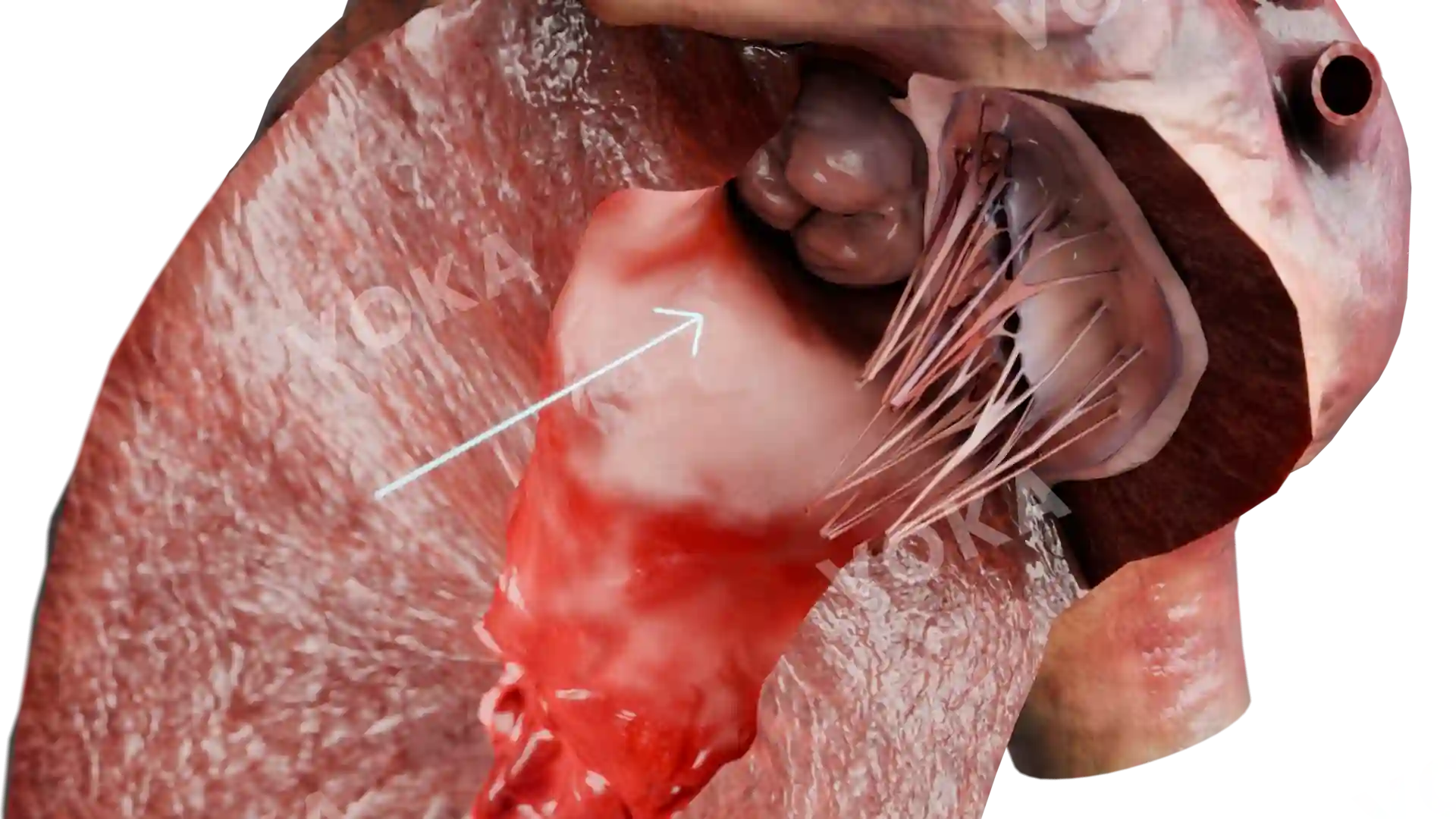
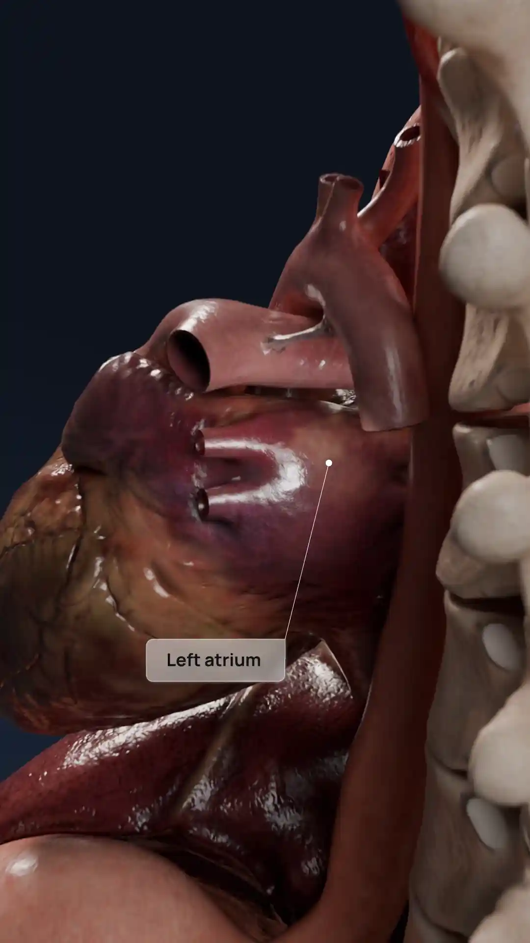
Flap of detached intima distal to the orifice of the left subclavian artery false and true lumens image - 30009
Cardiovascular system
Select license
More information
Details
Item successfully added to the cart