/Acute%20Otitis%20Media%20-%20Perforation%2C%202.webp)
/Acute%20Otitis%20Media%20-%20Perforation%2C%201.webp)


Description
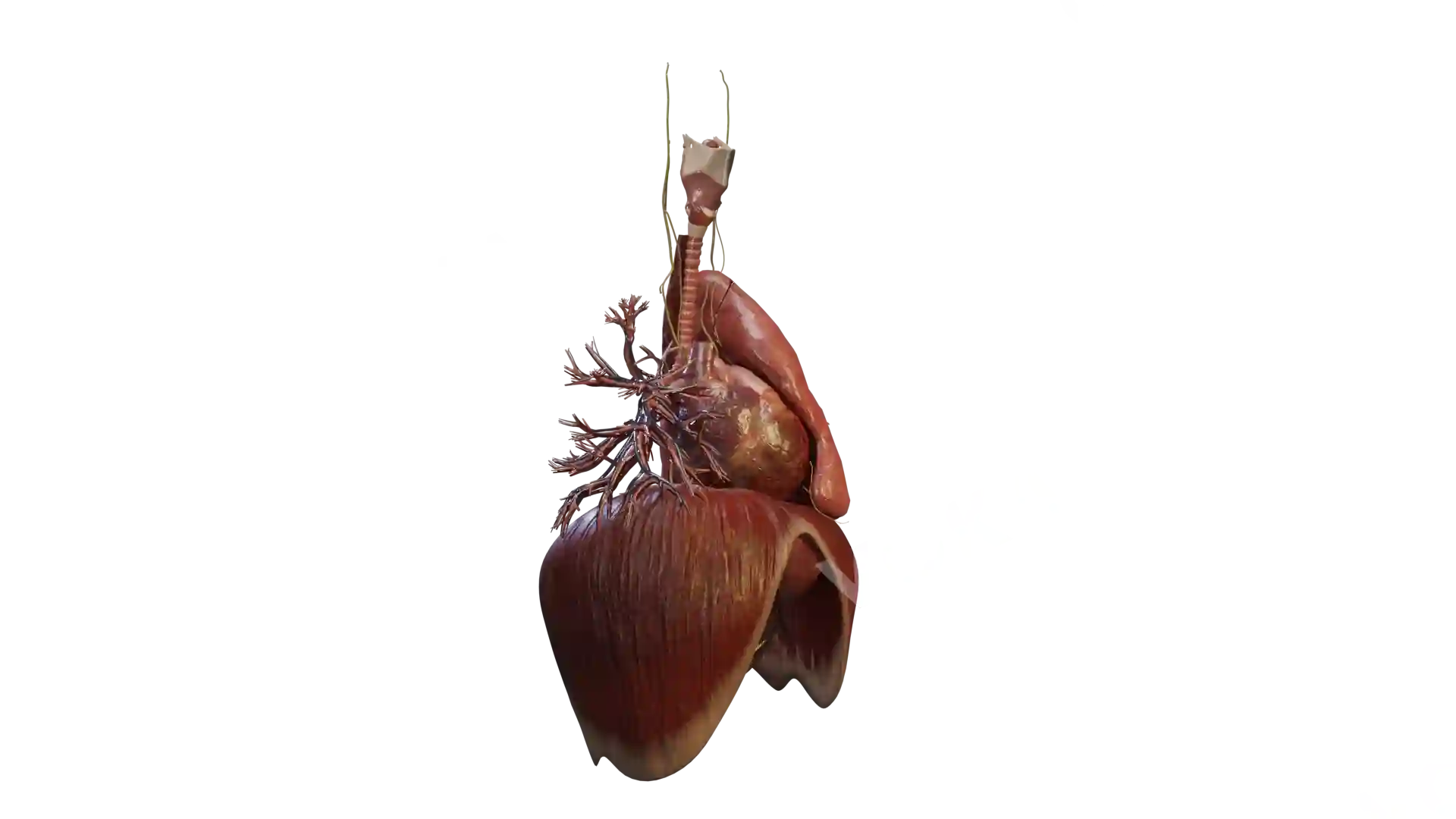
This detailed anatomical visualization showcases the human heart with a ventricular septal defect (VSD), highlighting its structural impact on cardiac function. The image captures the defect in the interventricular septum, allowing abnormal blood flow between the left and right ventricles. The surrounding structures, including the lungs, major blood vessels, and diaphragm, are clearly depicted to provide a comprehensive understanding of the condition. The image effectively demonstrates the increased workload on the heart due to the mixing of oxygenated and deoxygenated blood, emphasizing the clinical significance of VSD. This resource is ideal for medical education, patient counseling, and clinical reference.
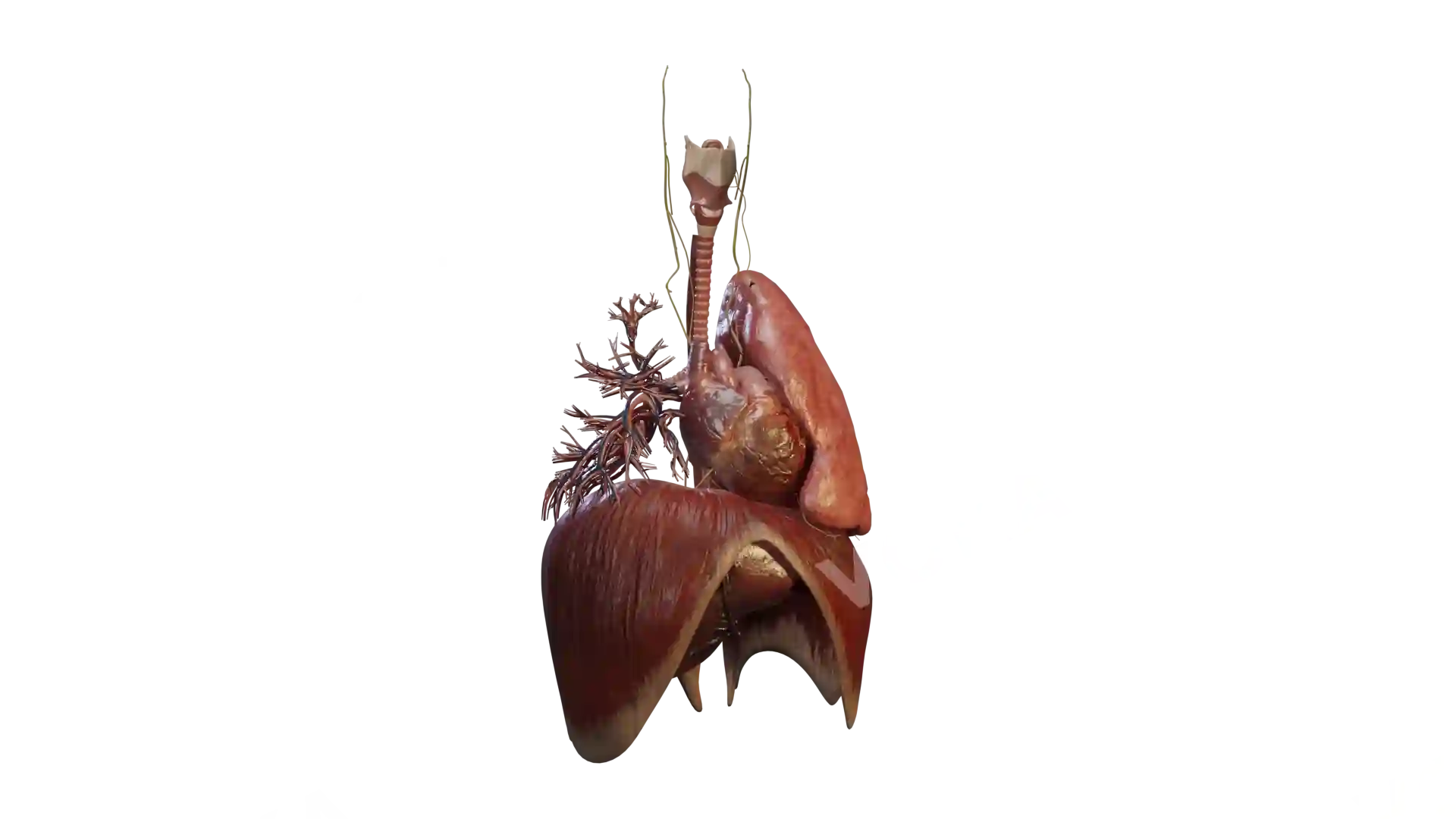
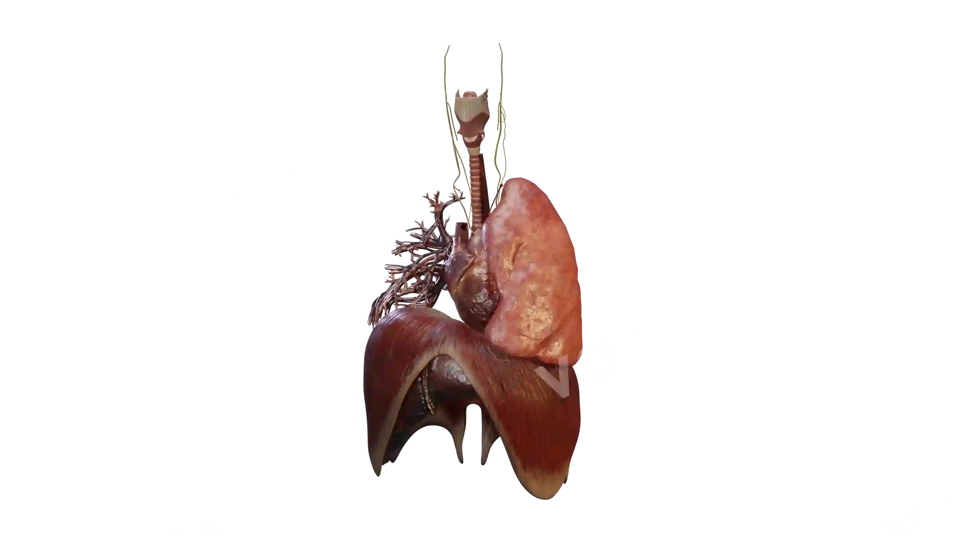
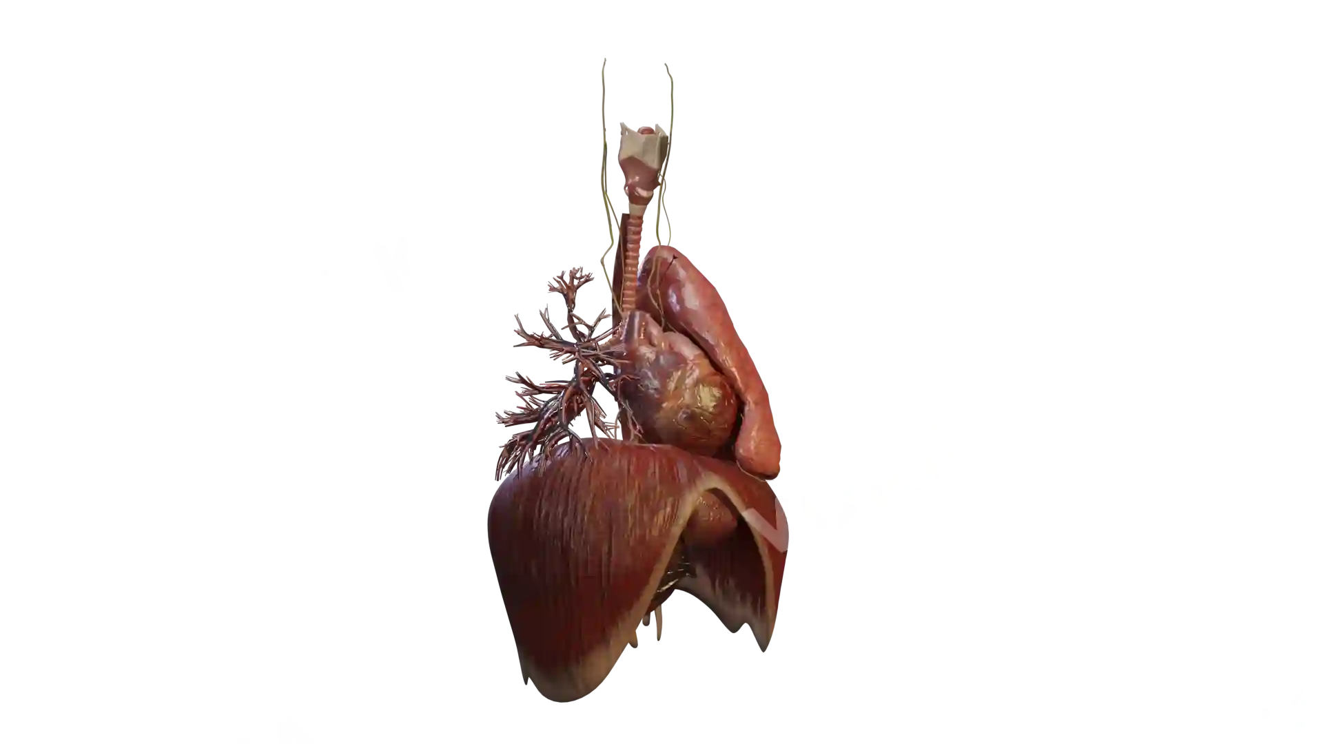
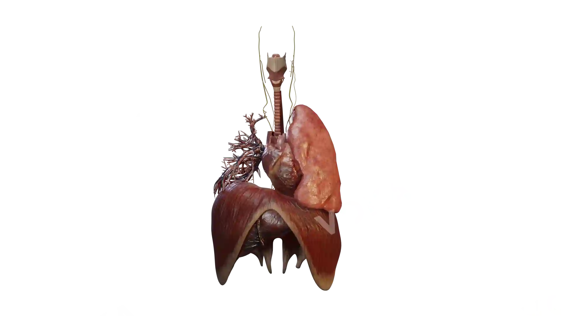
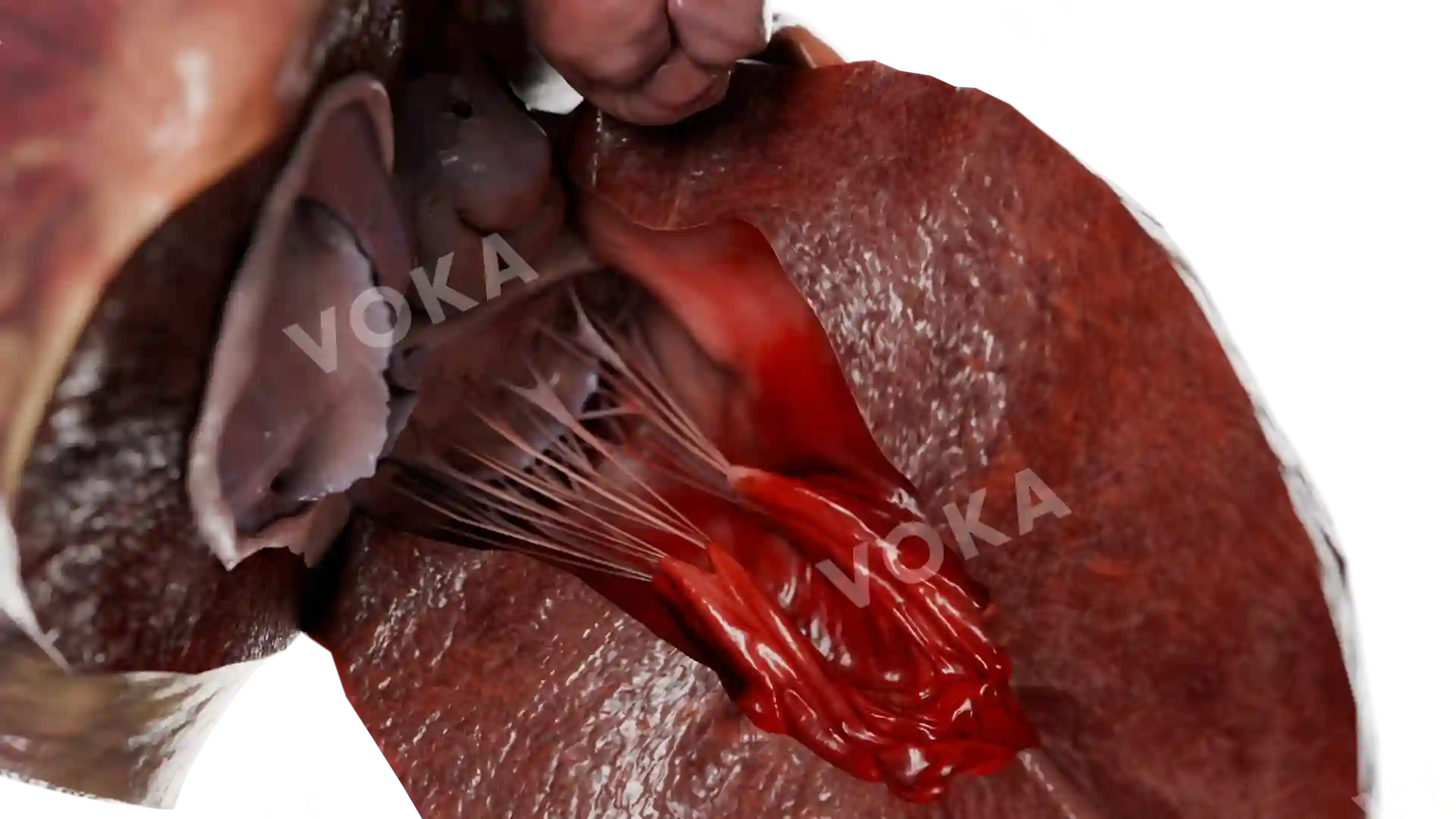
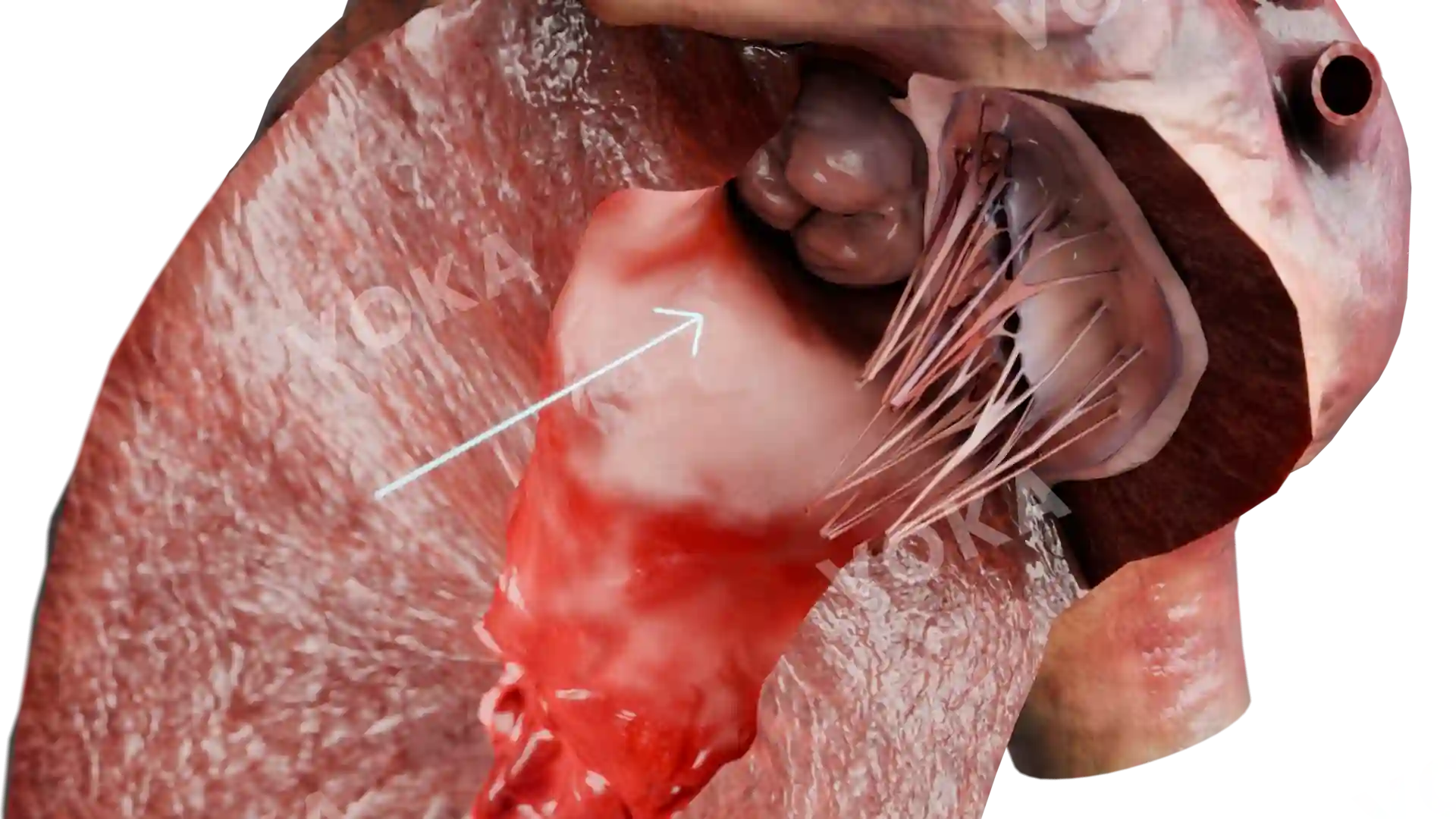
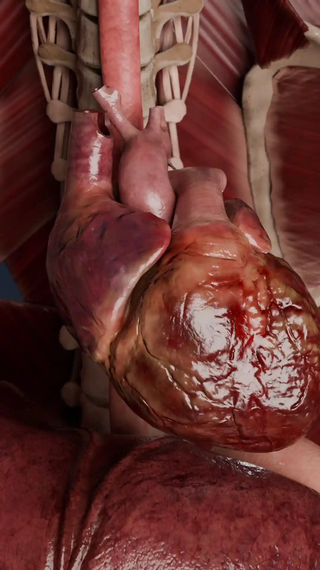
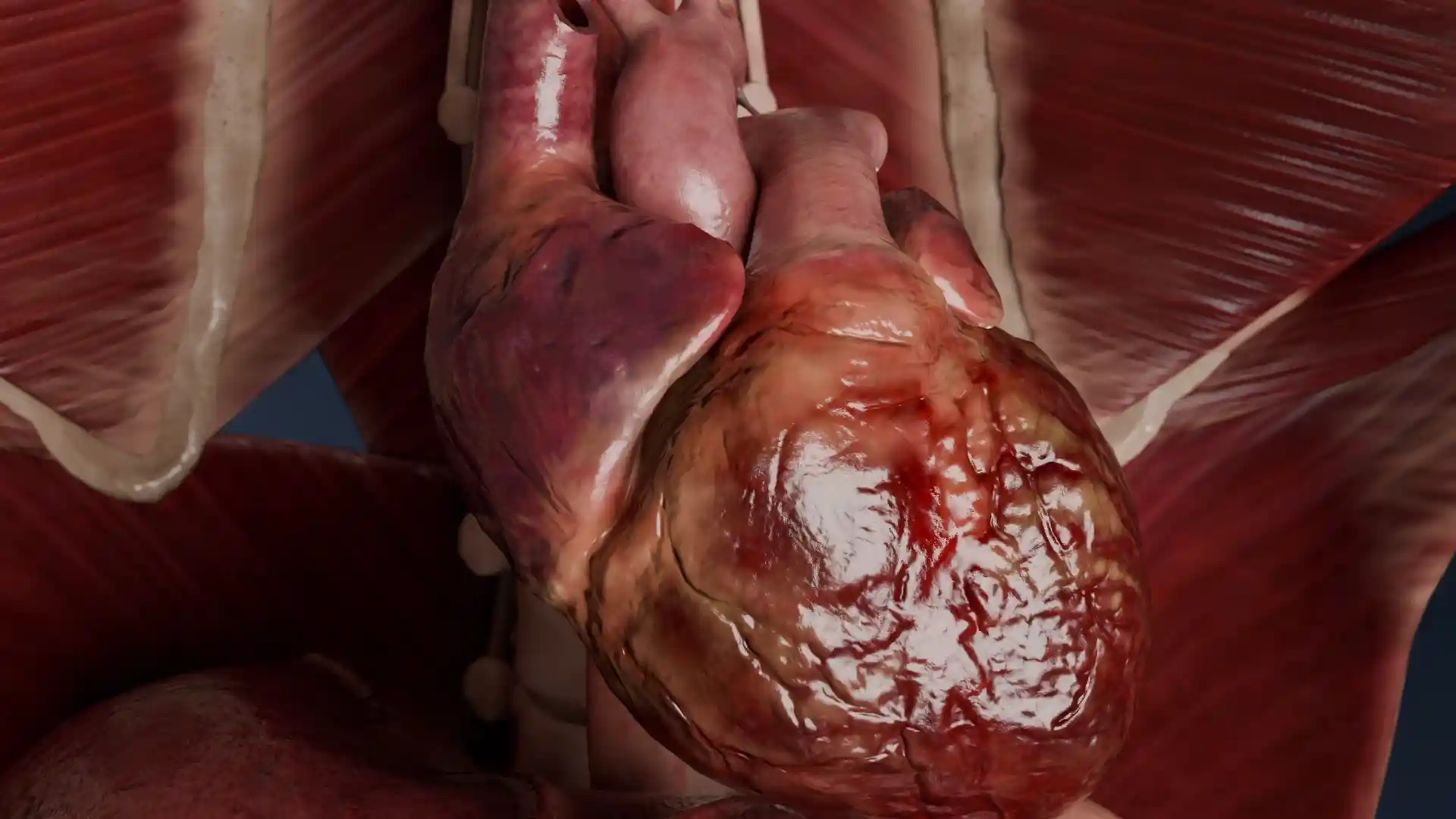
Heart with VSD and its surrounding structures image - 25014
Cardiovascular system
Select license
More information
Details
Item successfully added to the cart