/Acute%20Otitis%20Media%20-%20Perforation%2C%202.webp)
/Acute%20Otitis%20Media%20-%20Perforation%2C%201.webp)


Description
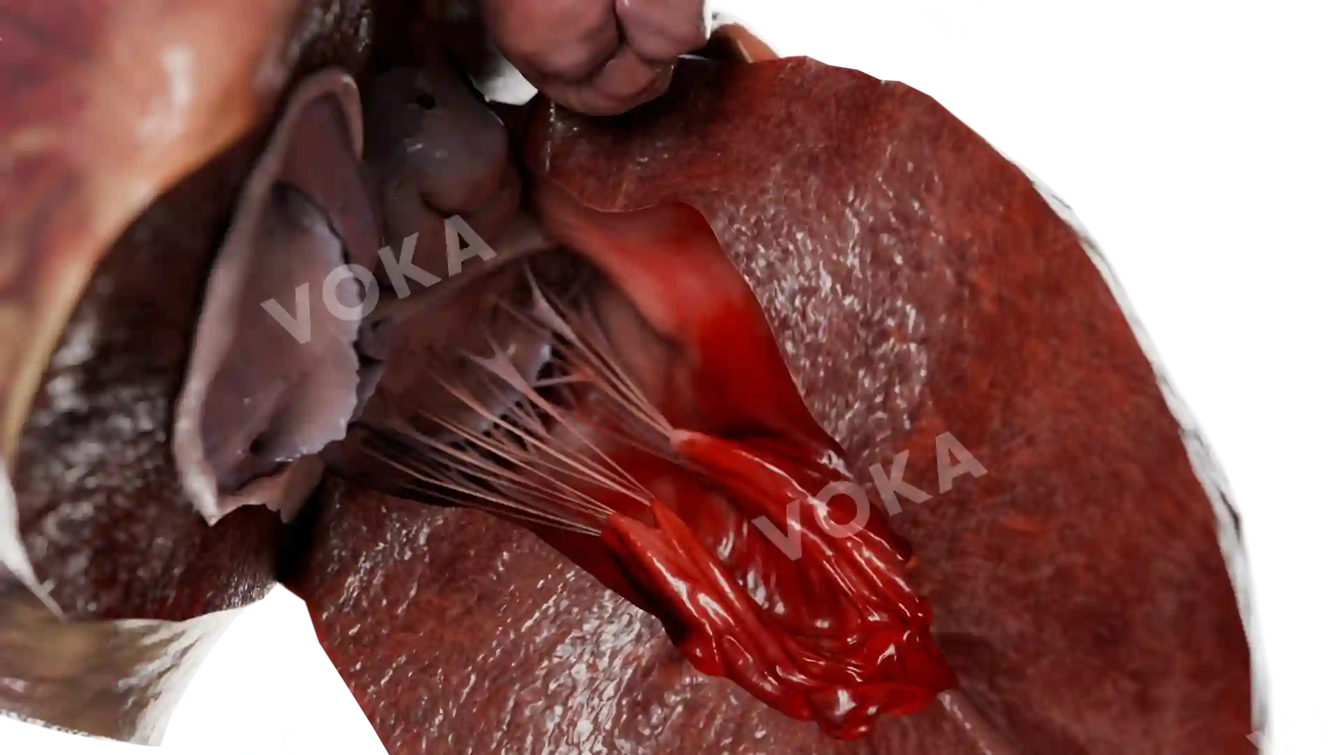
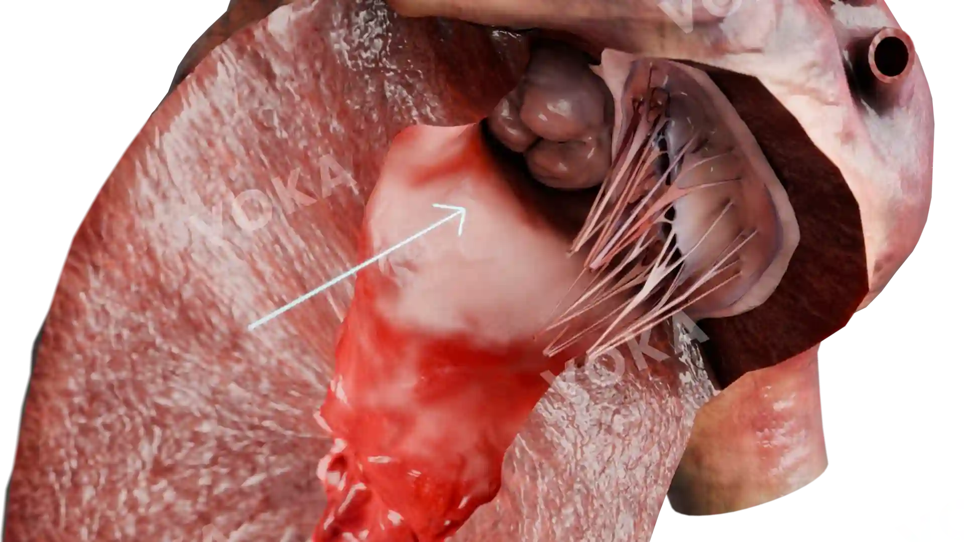
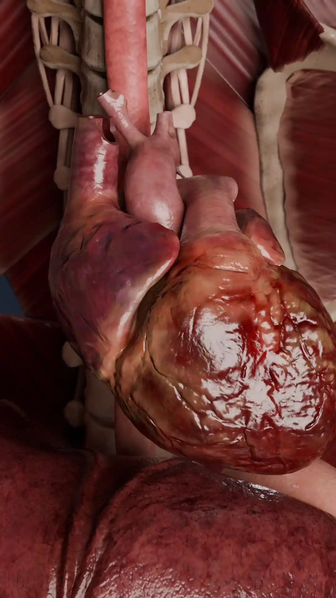
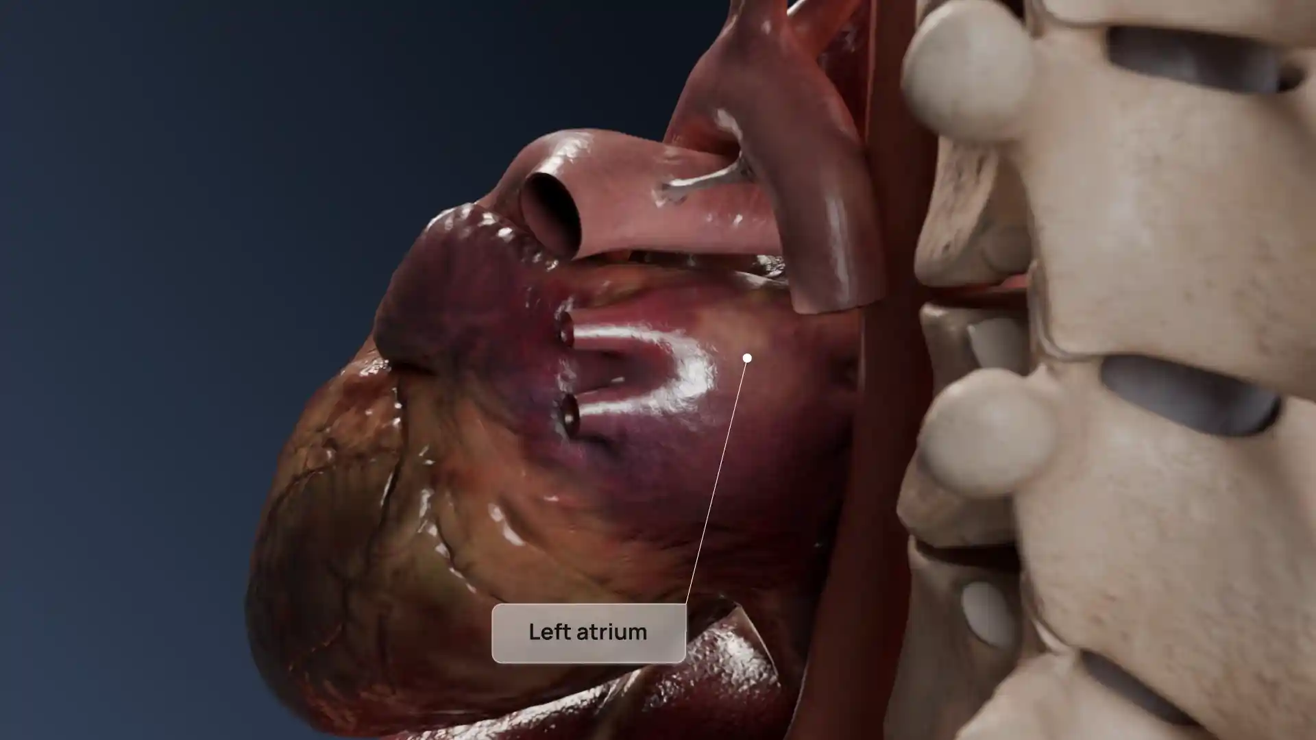
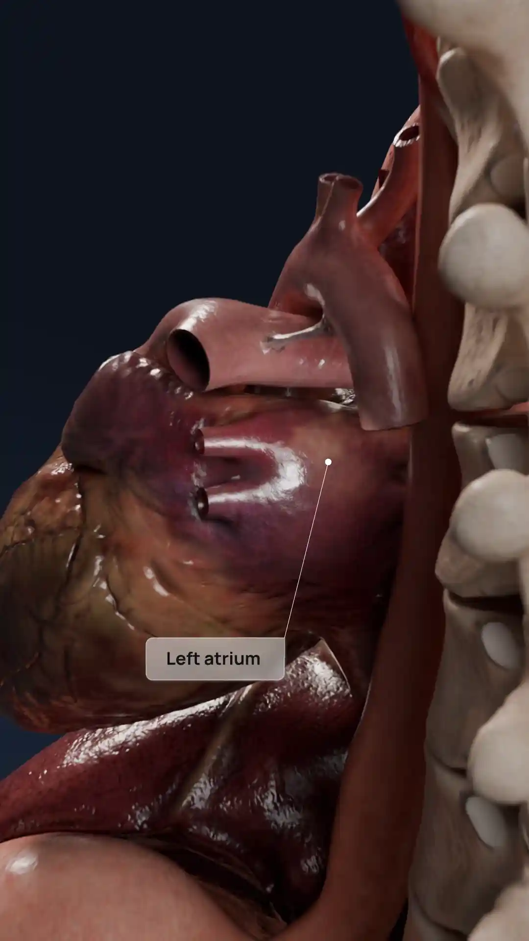
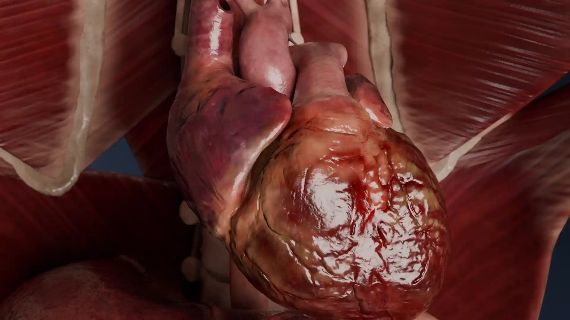
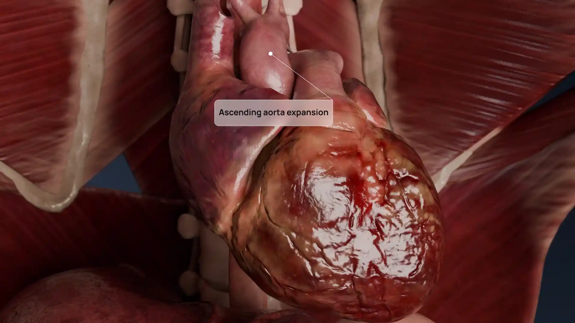
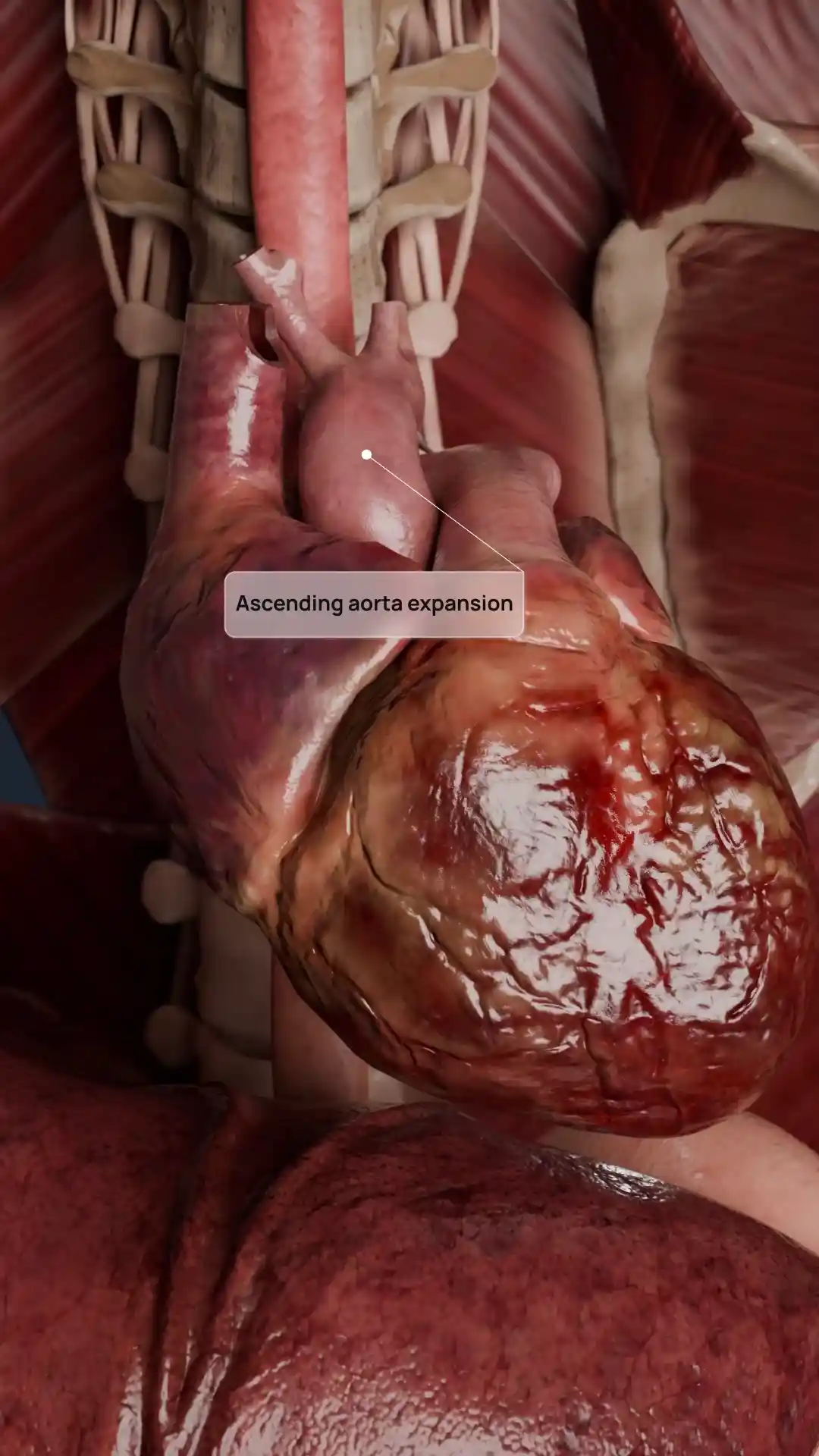
This detailed 3D image captures the structural abnormalities of rheumatic aortic valve stenosis, a condition often resulting from rheumatic fever. The image clearly shows the hallmark features, like thickened, fused valve leaflets and a narrowed orifice that obstructs blood flow from the left ventricle into the aorta. Such deformities significantly impair cardiac function and can lead to hypertrophy and heart failure if left untreated. This visualization provides a powerful tool for medical professionals and students to study the anatomical changes caused by chronic inflammation and scarring. It supports clinical education, diagnosis, surgical planning, and patient explanation with unmatched anatomical accuracy.
Rheumatic aortic valve stenosis image - 30105
Cardiovascular system
Select license
More information
Details
Item successfully added to the cart