/Acute%20Otitis%20Media%20-%20Perforation%2C%202.webp)
/Acute%20Otitis%20Media%20-%20Perforation%2C%201.webp)


Description
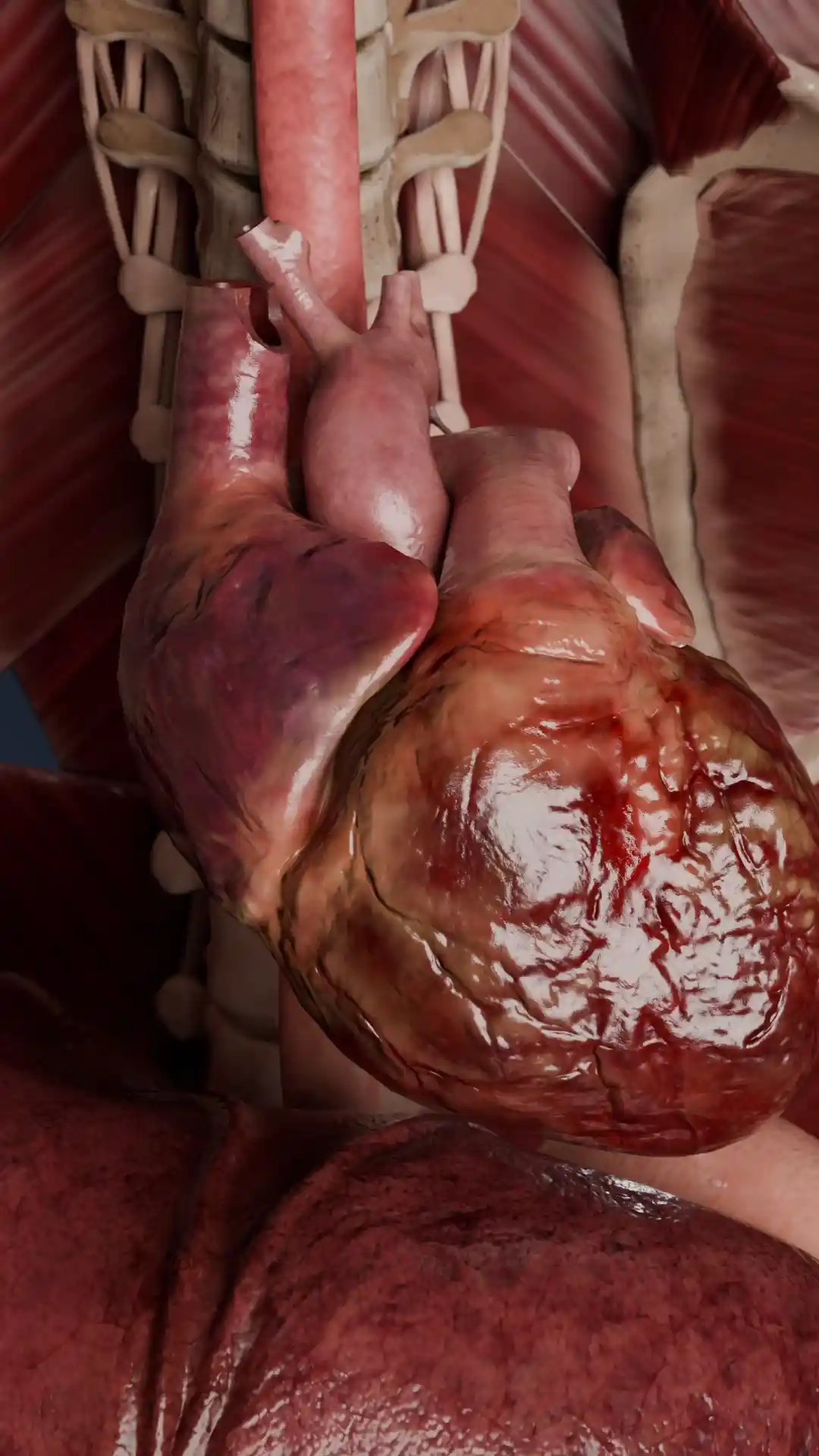
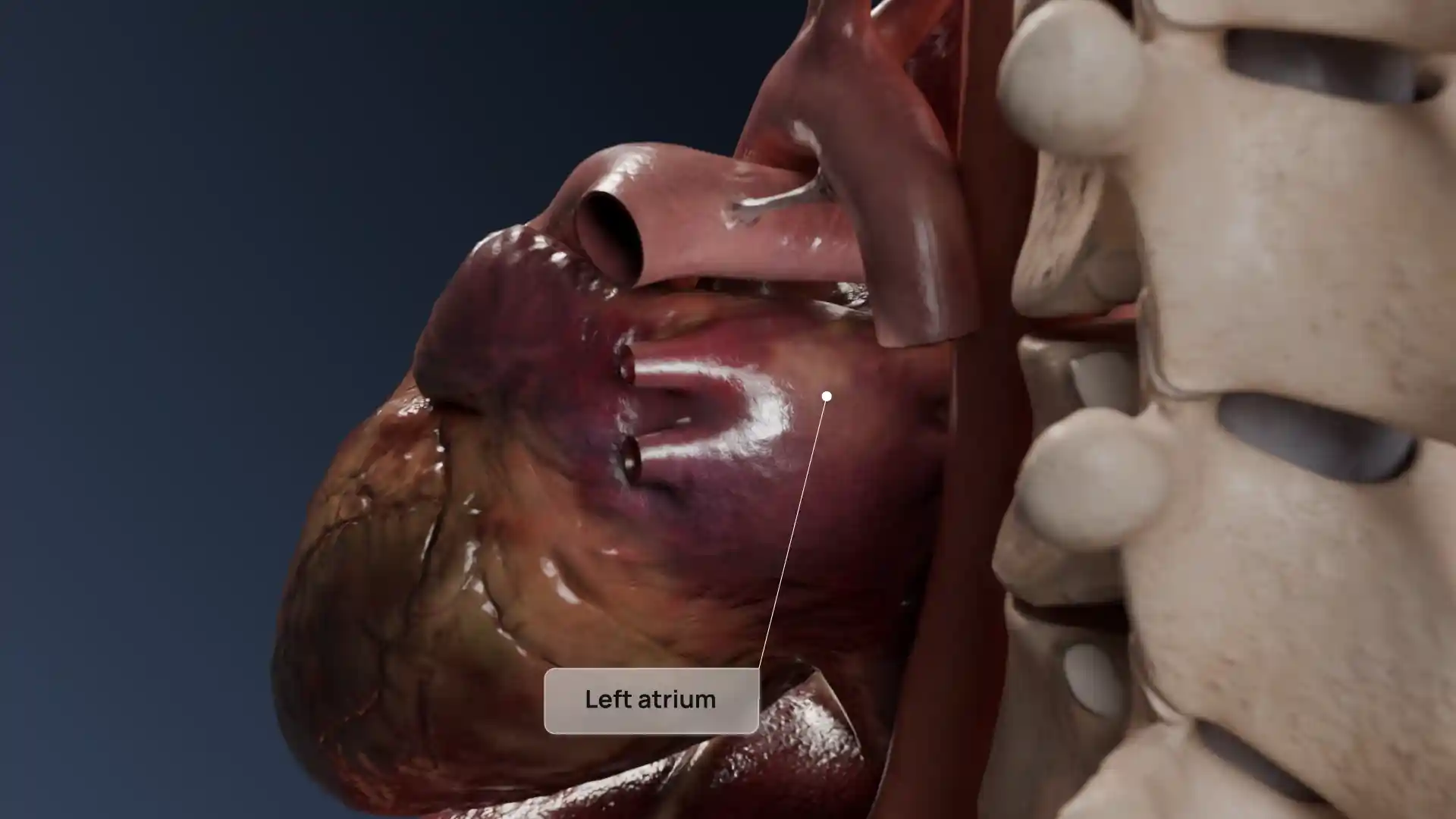
This high-resolution 3D image illustrates thickened and fused tricuspid valve leaflets, a hallmark of tricuspid stenosis. The constricted valve opening limits blood flow from the right atrium to the right ventricle, causing increased atrial pressure and systemic venous congestion. The visualization captures the anatomical distortion in detail, from the fibrotic, immobile leaflets to the narrowed orifice. Such pathologies are often linked to rheumatic heart disease or congenital abnormalities. Ideal for medical students, cardiologists, and educators, this model offers a precise and realistic view of tricuspid stenosis to support clinical learning, diagnostic practice, surgical planning, and patient communication.
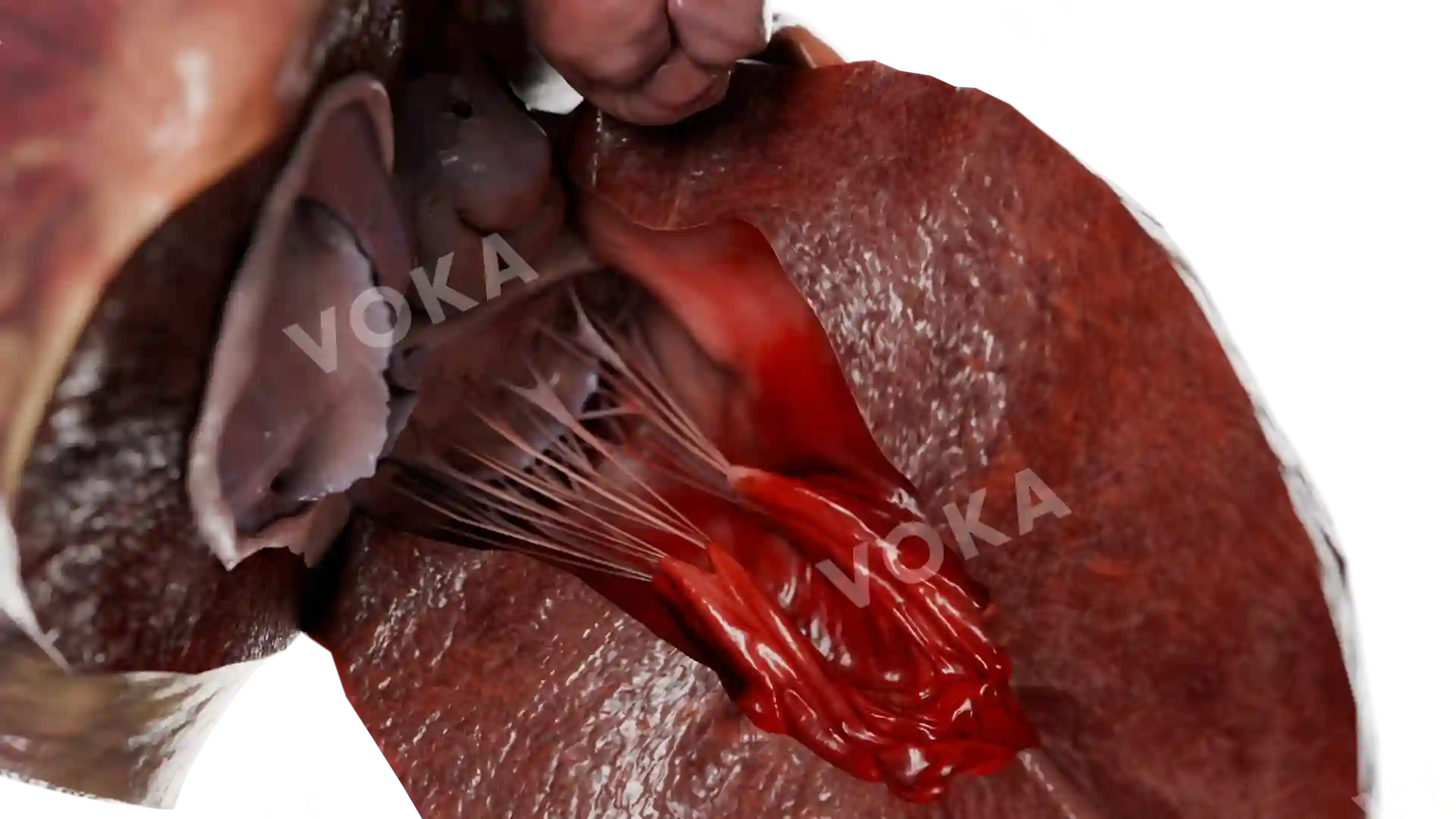
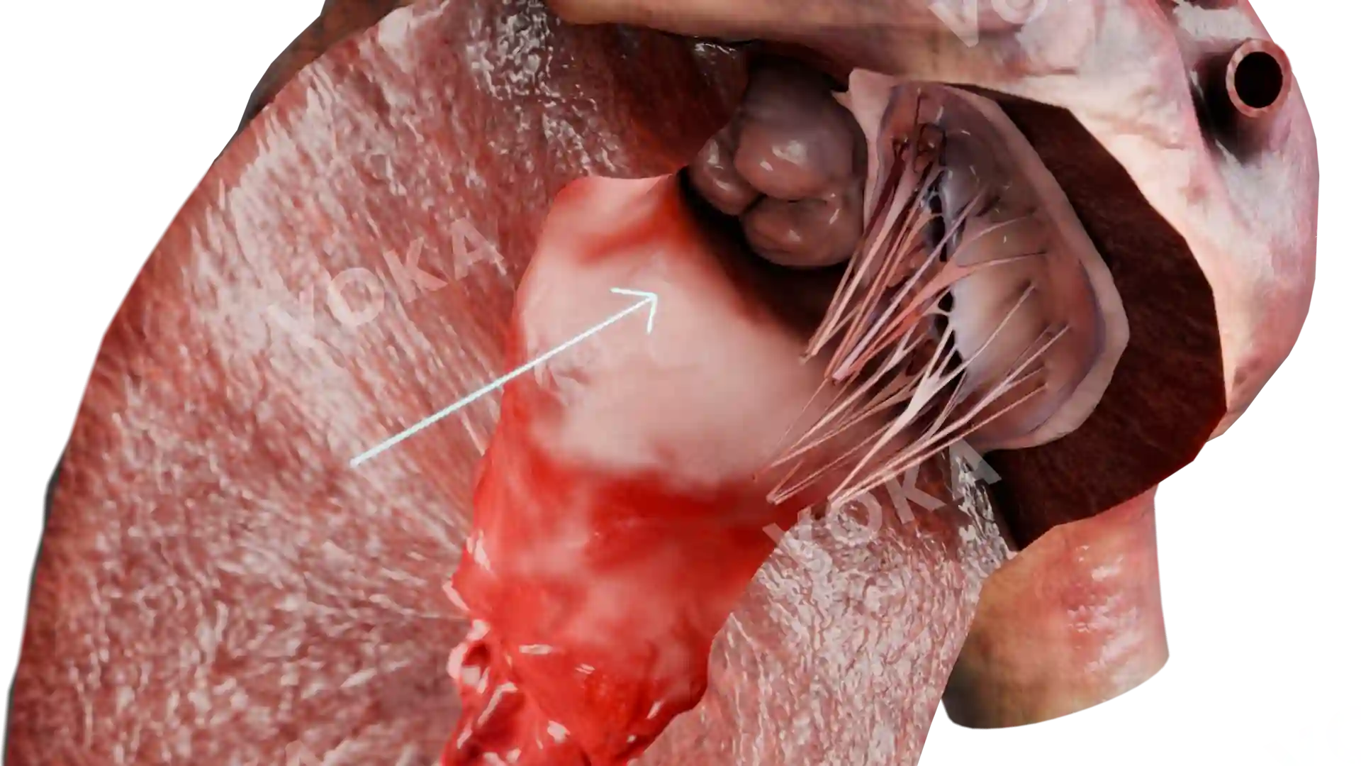
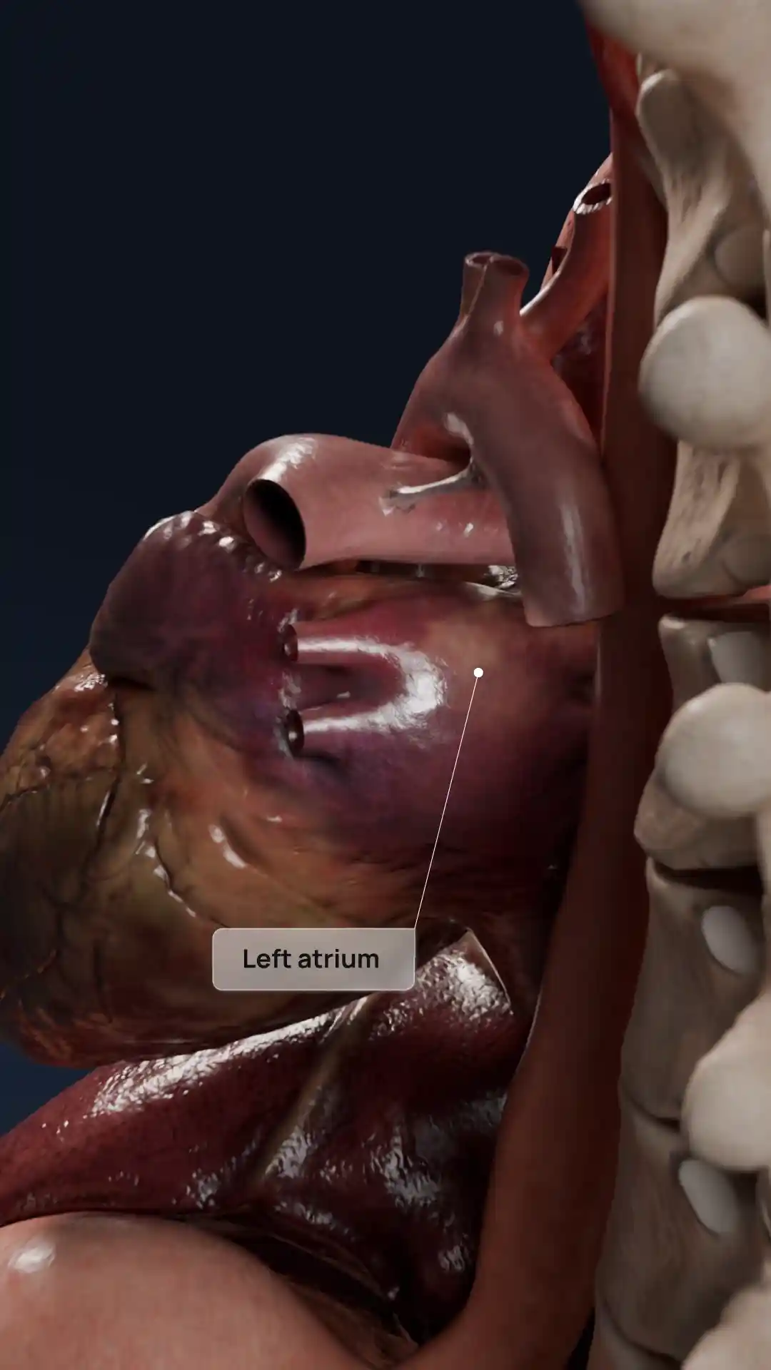
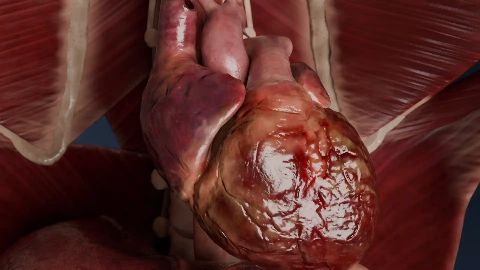
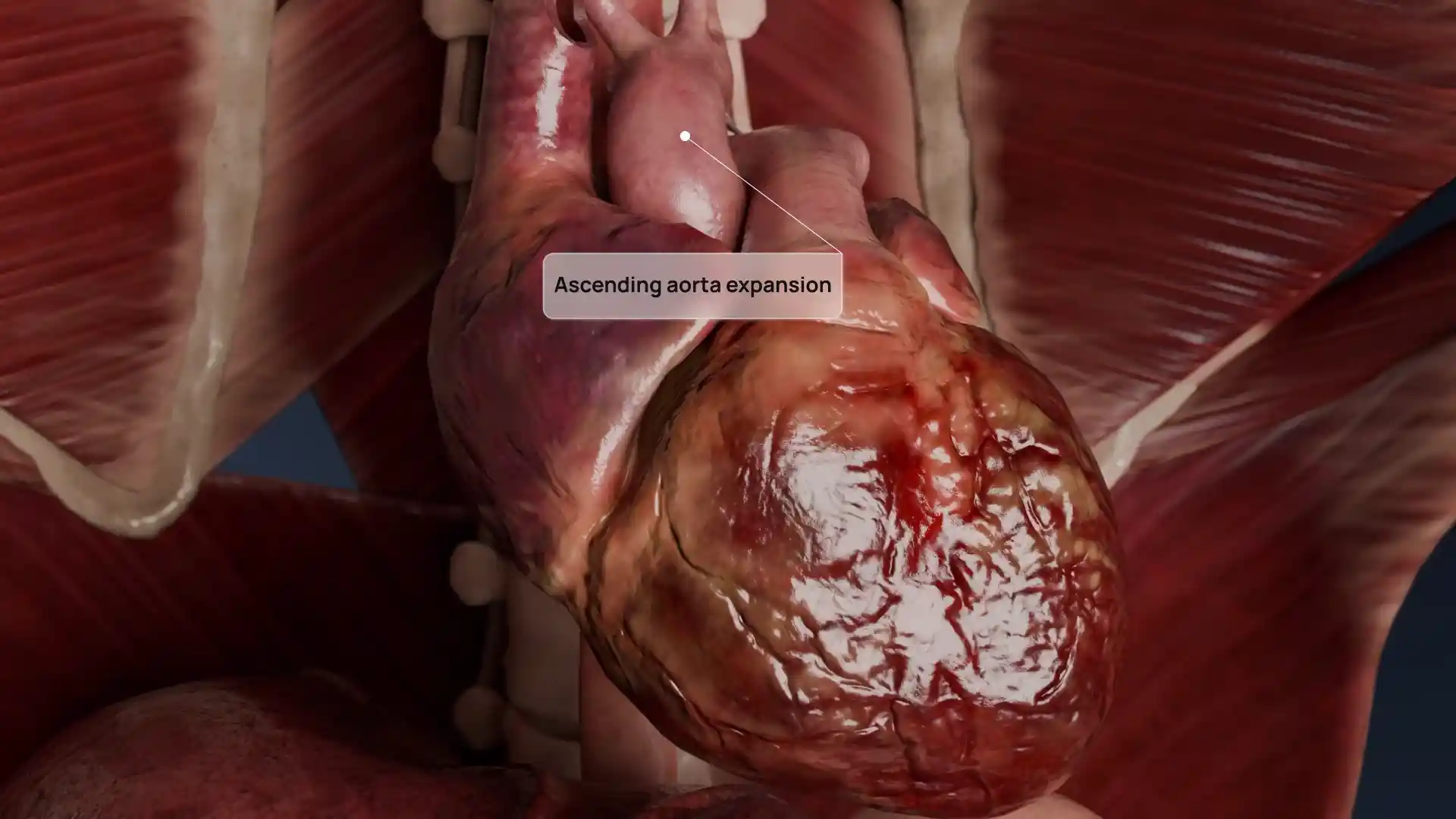
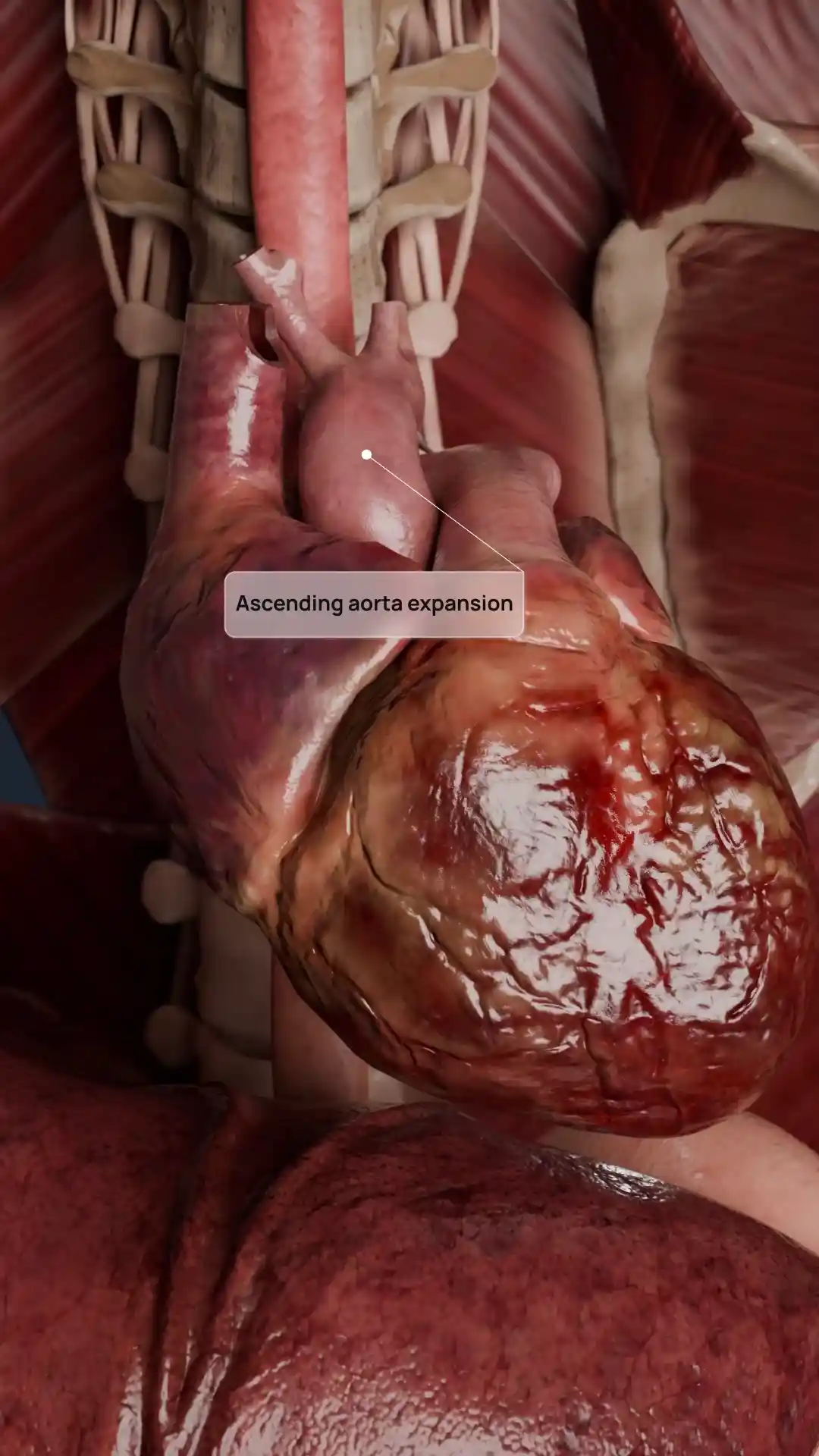
Thickened and fused tricuspid valve leaflets in tricuspid stenosis image - 30106
Cardiovascular system
Select license
More information
Details
Item successfully added to the cart