/Acute%20Otitis%20Media%20-%20Perforation%2C%202.webp)
/Acute%20Otitis%20Media%20-%20Perforation%2C%201.webp)


Description
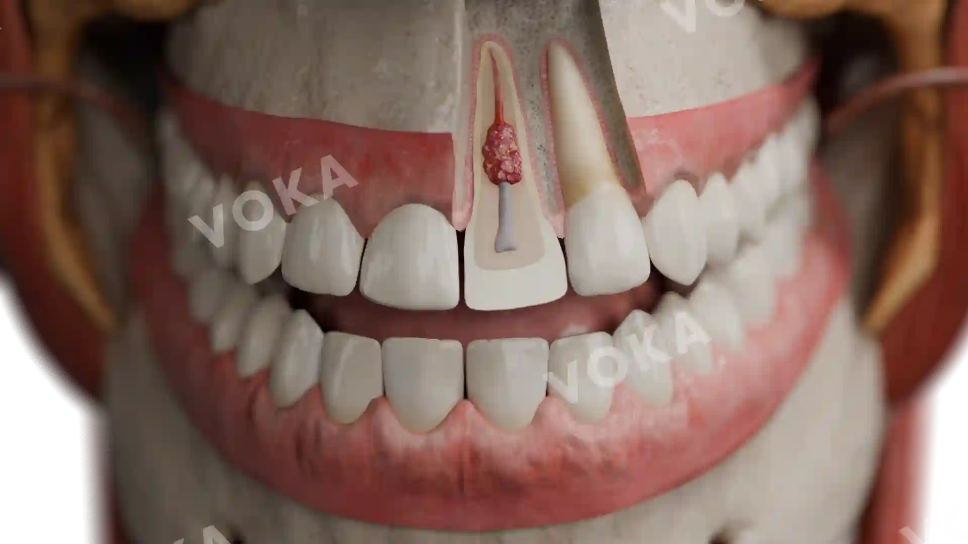
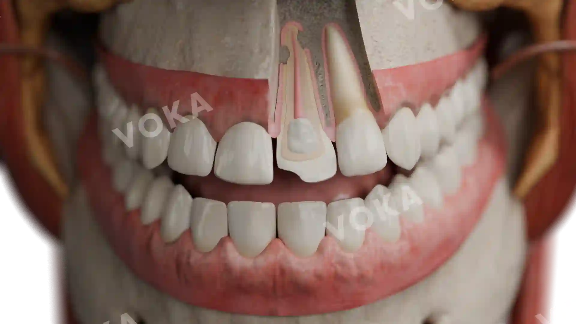
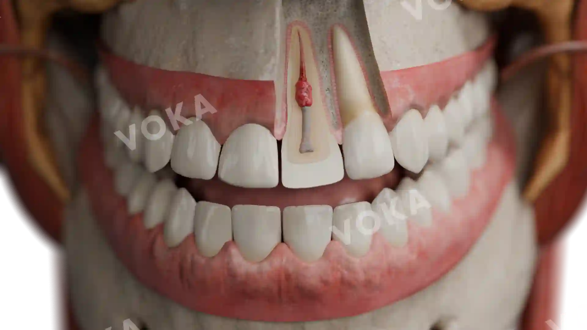
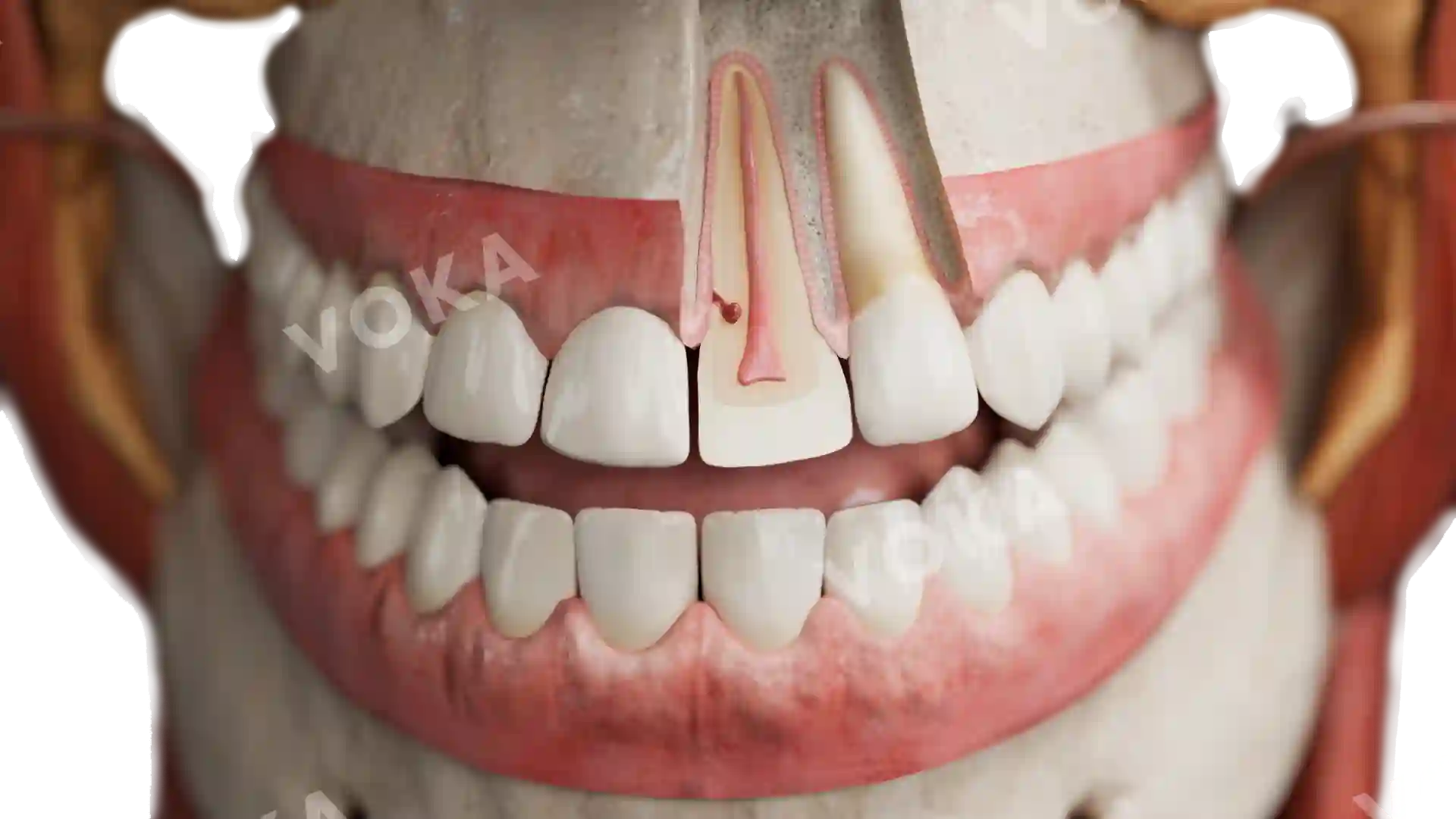
This comprehensive anatomical image highlights the presentation of Amelogenesis Imperfecta, hypomaturic type, within the broader context of facial anatomy. The teeth display hallmark signs of hypomaturic enamel defects, including a patchy, discolored appearance with varying shades of yellow and brown. The enamel appears thin, rough, and uneven, with some areas showing signs of surface degradation and reduced translucency. The image uniquely integrates dental structures with surrounding facial muscles, blood vessels, and nerves, offering a holistic view of how this condition fits within overall craniofacial anatomy. This high-resolution illustration is a valuable resource for dental students, healthcare professionals, and educators, perfect for dental pathology courses, anatomy classes, and clinical presentations.
Related items


Amelogenesis imperfecta, hypoplastic image - 25022
Dentistry
Select license
More information
Details
Item successfully added to the cart