/Acute%20Otitis%20Media%20-%20Perforation%2C%202.webp)
/Acute%20Otitis%20Media%20-%20Perforation%2C%201.webp)


Description
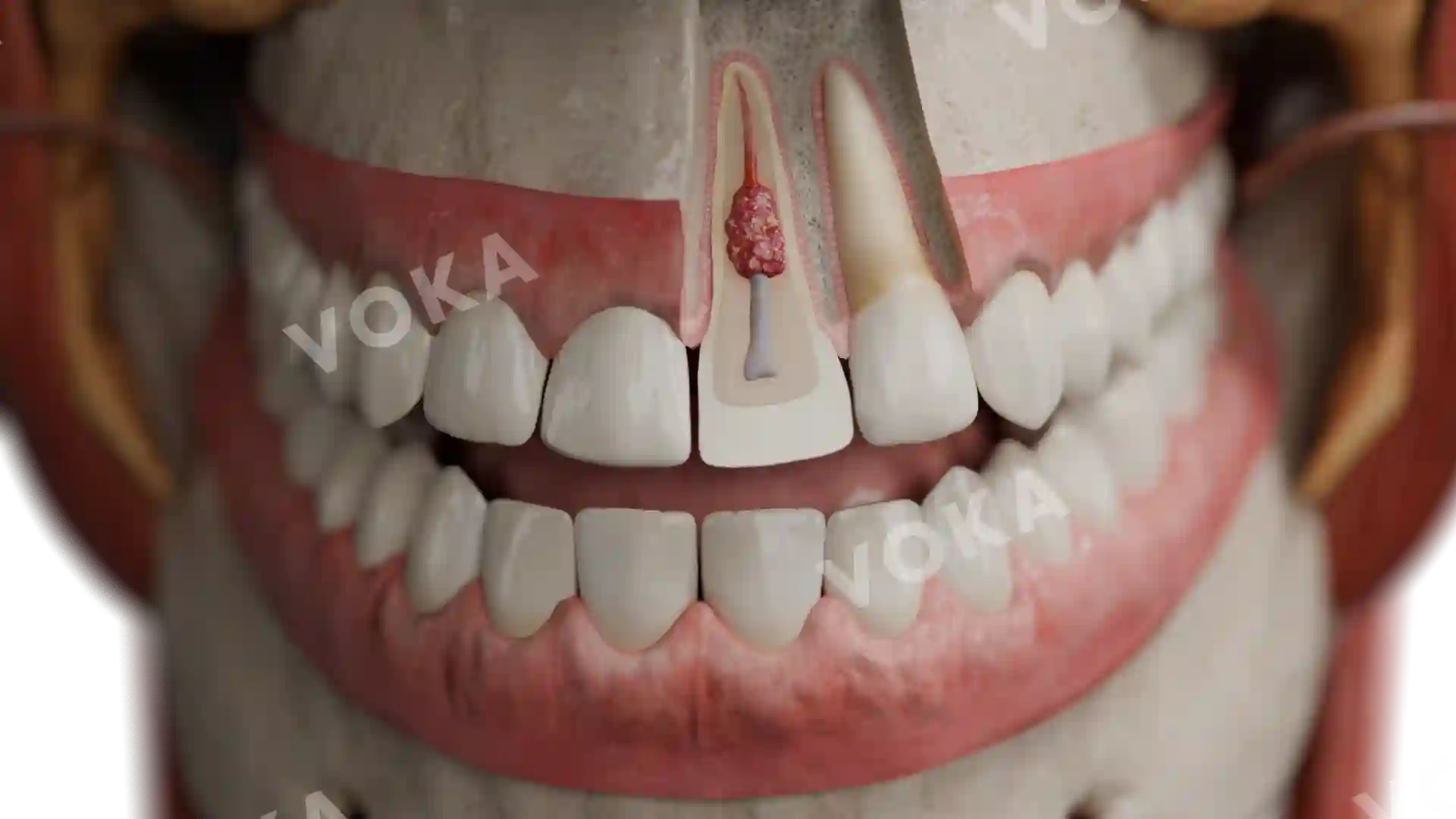
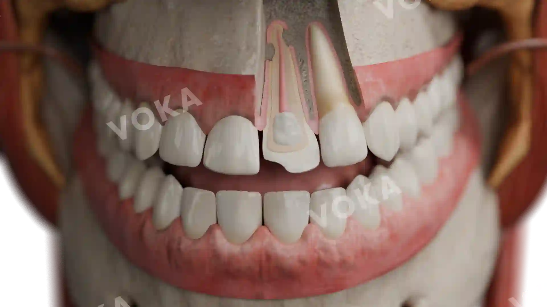
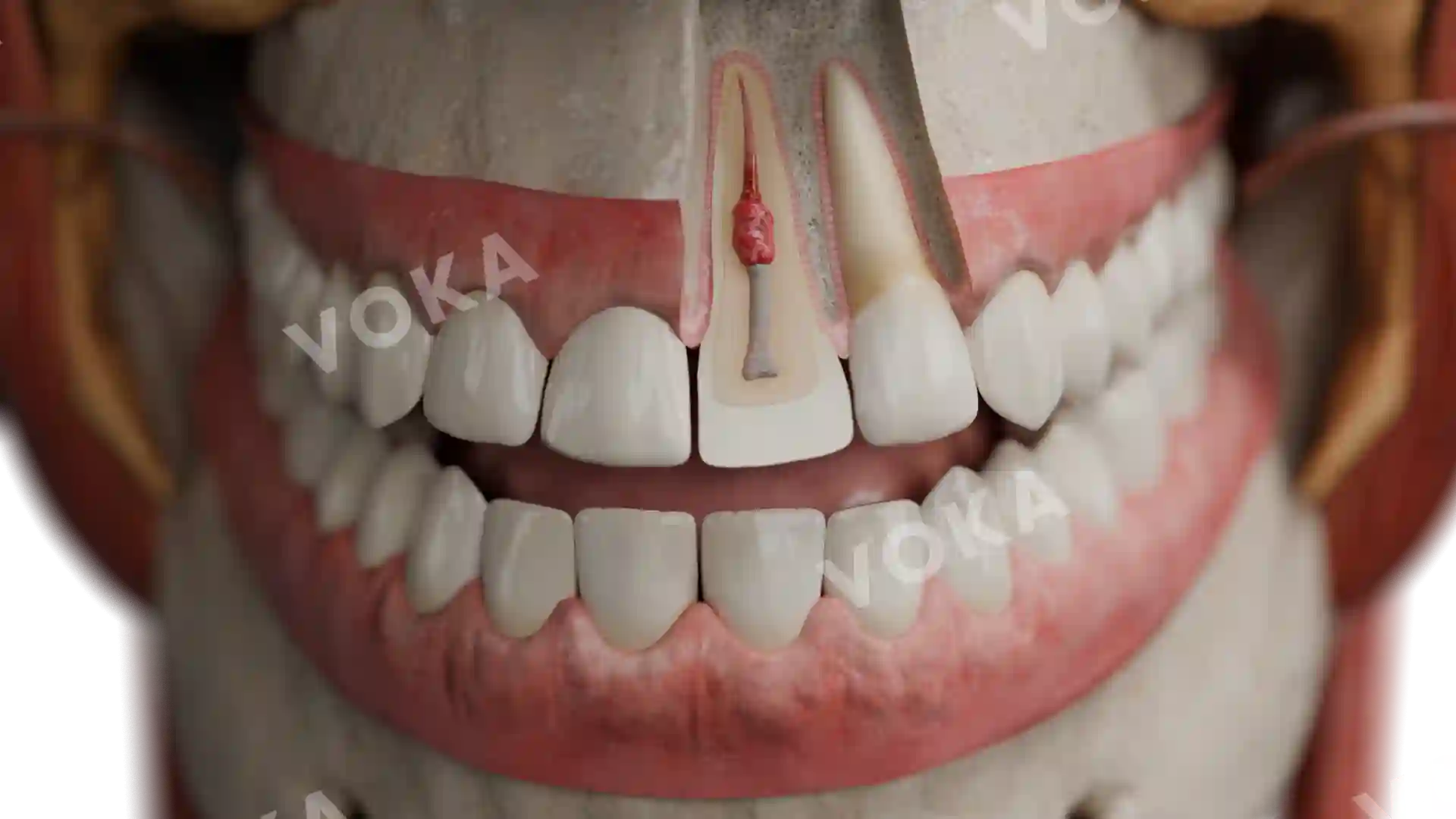
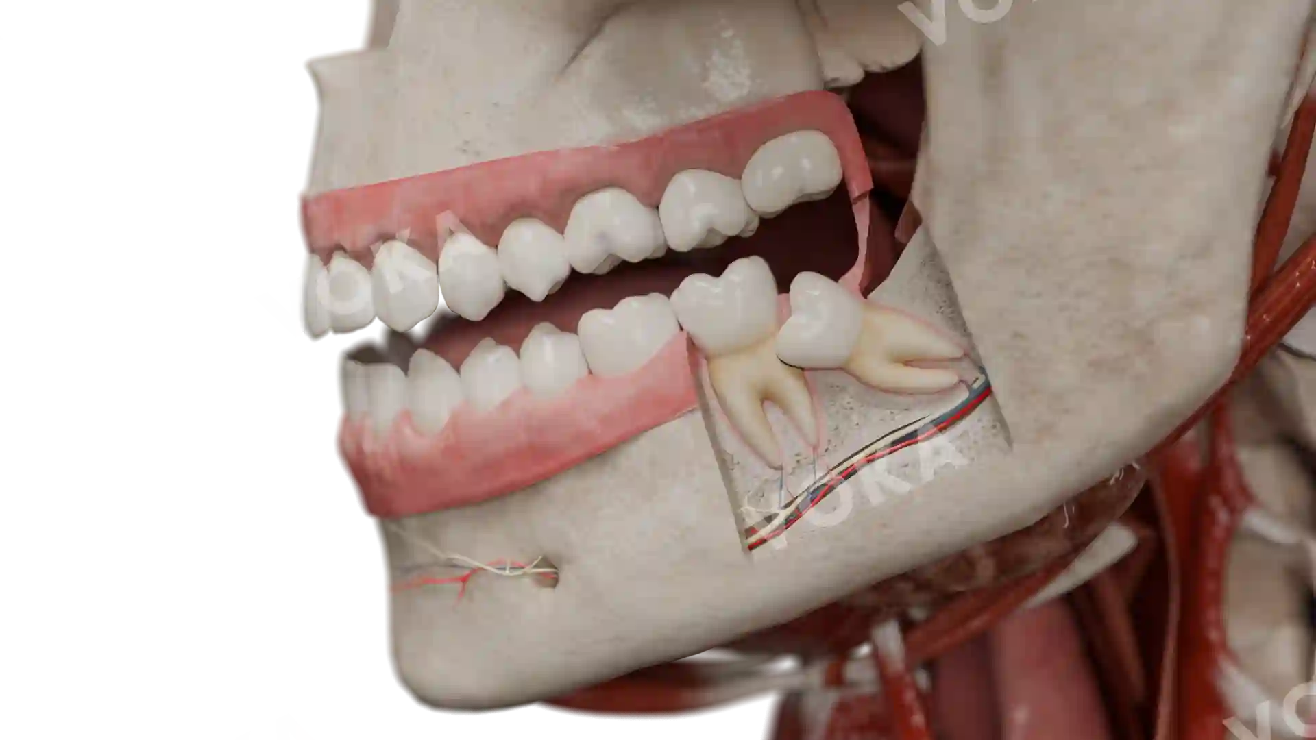
This detailed anatomical illustration showcases Amelogenesis Imperfecta, hypomaturic type, with a focus on both dental and facial structures. The teeth exhibit typical signs of hypomaturic enamel defects, including uneven discoloration with yellowish-brown hues and a rough, chalky texture. The image also features an in-depth view of the surrounding facial anatomy, highlighting the muscles, nerves, and vascular networks that support oral and facial function. The symmetrical presentation of the face, combined with detailed exposure of internal structures, provides a comprehensive understanding of the condition within the context of craniofacial anatomy. This high-resolution image serves as an excellent educational tool for dental students, healthcare professionals, and educators, offering valuable insights for anatomy courses, dental pathology studies, and clinical presentations.
Related items


Amelogenesis imperfecta, hypoplastic image - 25023
Dentistry
Select license
More information
Details
Item successfully added to the cart