/Acute%20Otitis%20Media%20-%20Perforation%2C%202.webp)
/Acute%20Otitis%20Media%20-%20Perforation%2C%201.webp)


_with_the_oral_cavity.webp)
Description
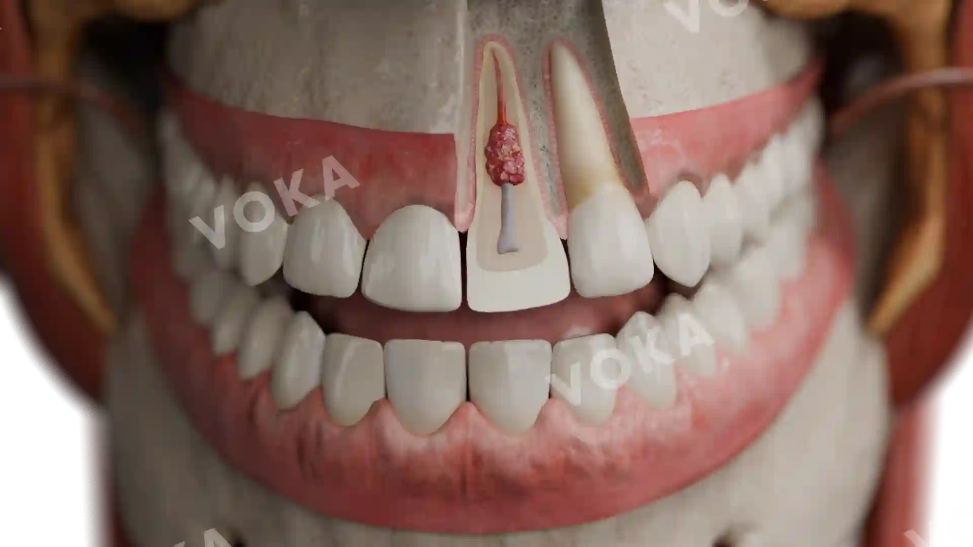
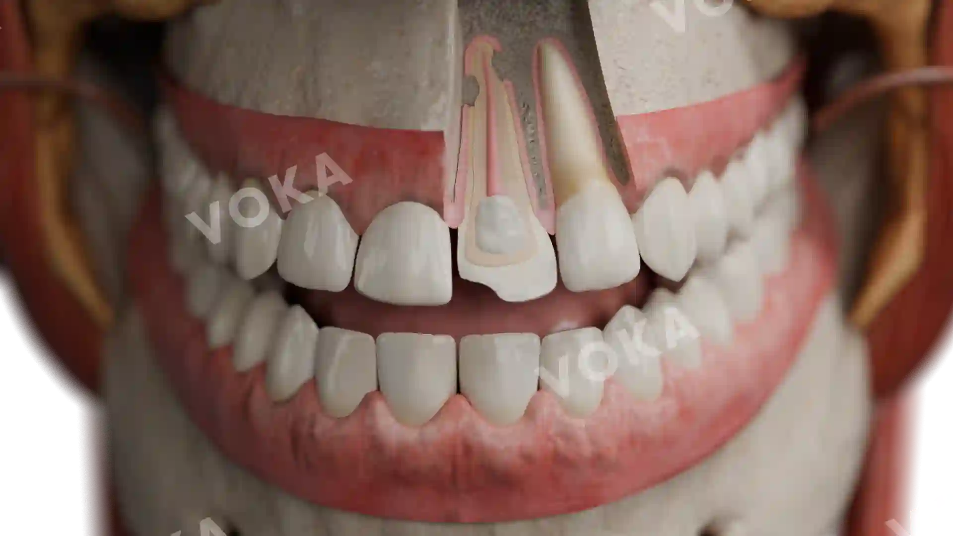
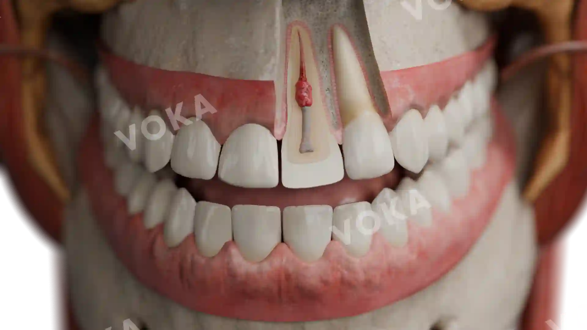
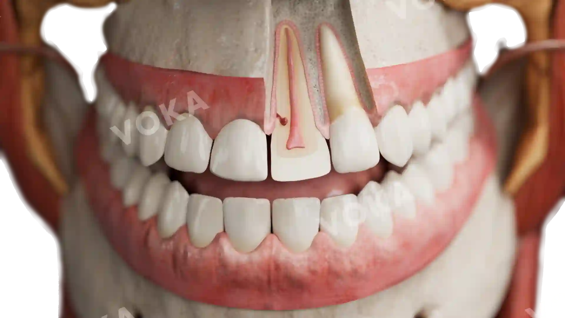
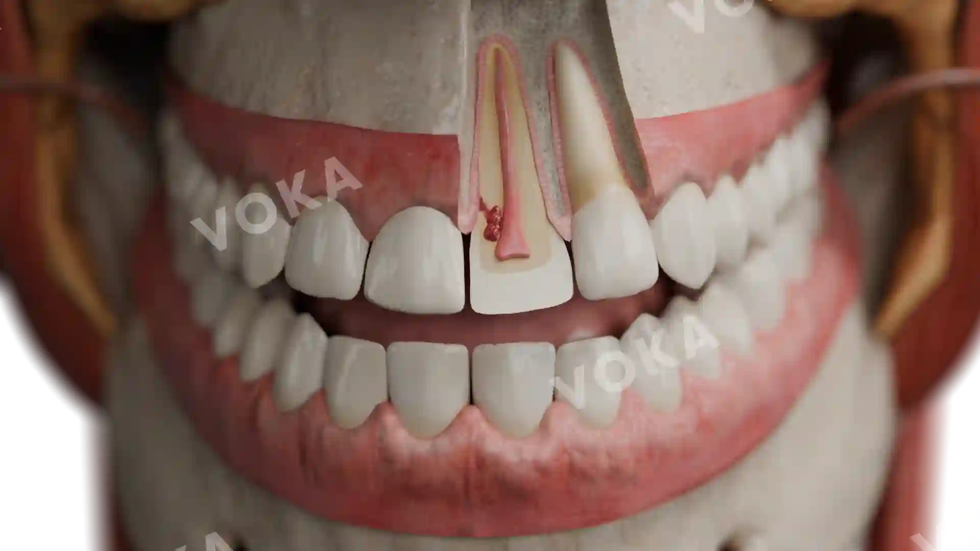
This anatomical illustration demonstrates a chronic apical abscess in a lower molar, with a clearly visible fistulous tract opening into the oral cavity. The sagittal section reveals the decayed crown and root filled with necrotic material, while the periapical region is enlarged and encapsulated by a pale, thick-walled inflammatory lesion. The abscess cavity is more organized than in acute cases, with a defined channel extending through the alveolar bone and soft tissue, indicating ongoing drainage. The gingiva and surrounding tissues appear slightly elevated, reflecting the persistent nature of the lesion. This image offers a detailed view of the chronic inflammatory process and its communication pathway to the oral surface.
Related items


Anatomy of a chronic apical abscess with communication (fistula) with the oral cavity image - 30068
Dentistry
Select license
More information
Details
Item successfully added to the cart