Anatomy of an acute apical abscess image

After purchasing you’ll get a link to a high-resolution file. Your image or video won’t contain watermarks.

Description

This detailed anatomical illustration depicts an acute apical abscess affecting a lower molar, presented in a sagittal section. The image clearly shows a necrotic pulp chamber filled with darkened, decayed tissue extending through the root canal. The abscess cavity appears slightly elevated, with visible compression of nearby structures, and the cortical plate is beginning to thin. This visual effectively captures the acute stage of infection with clear contrast between healthy, inflamed, and purulent regions.

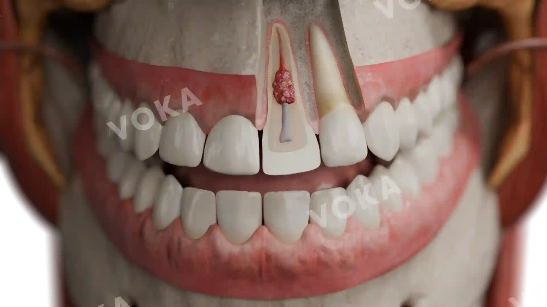
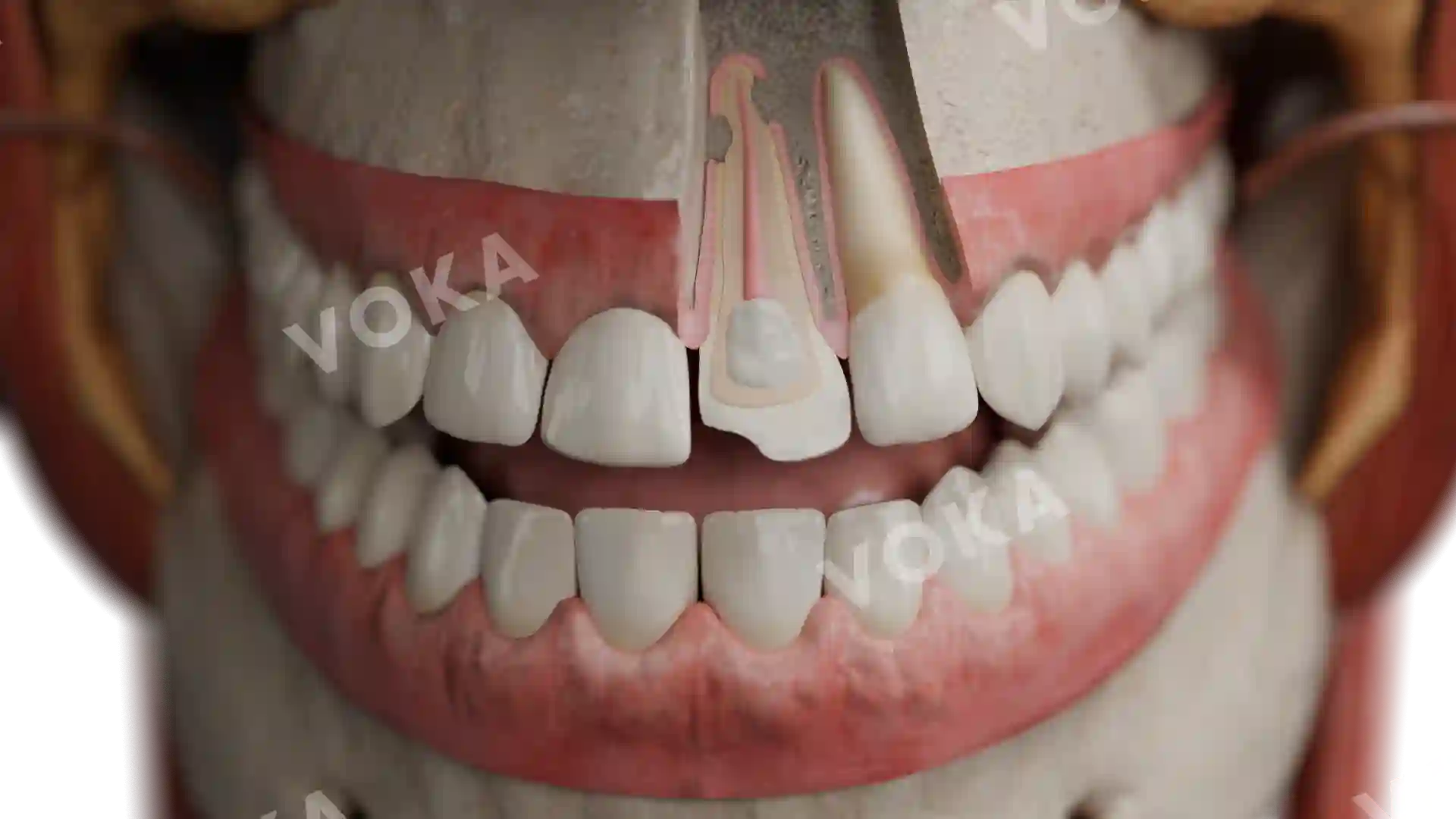
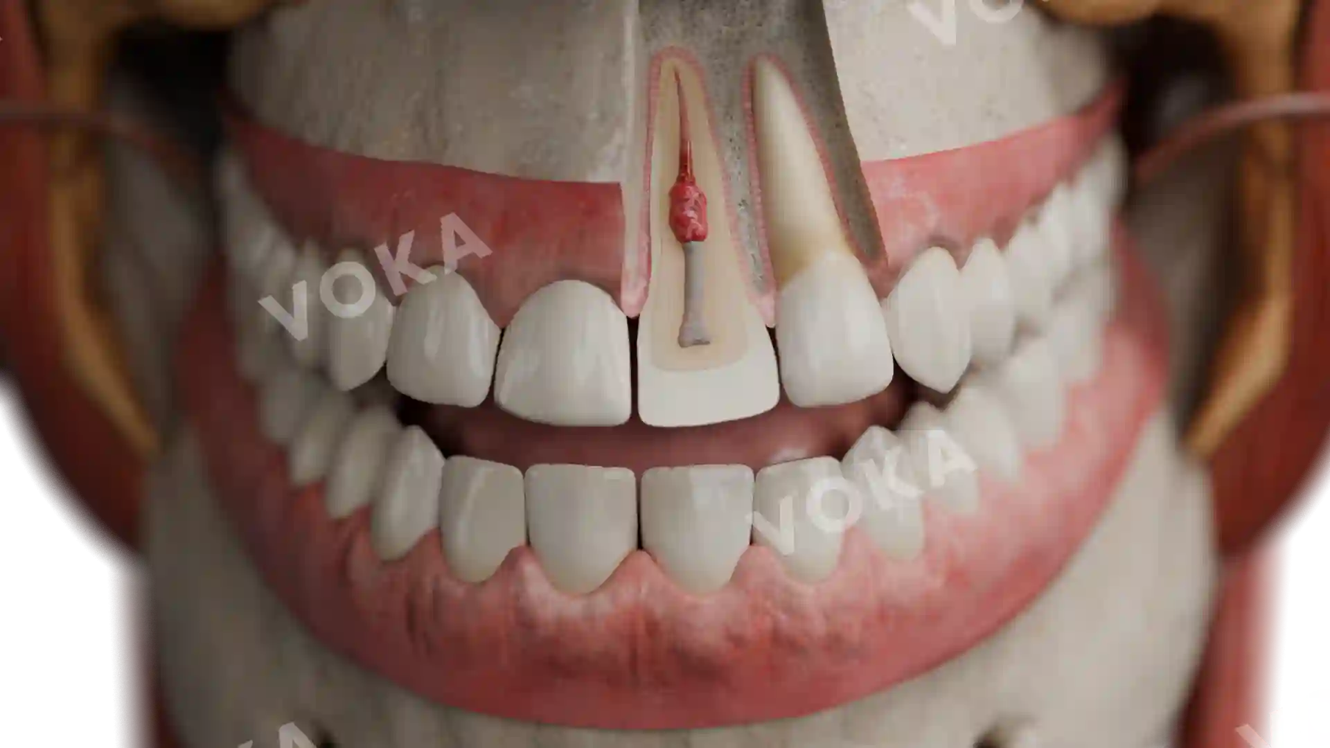
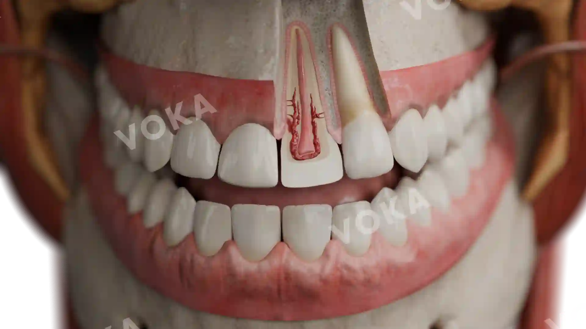
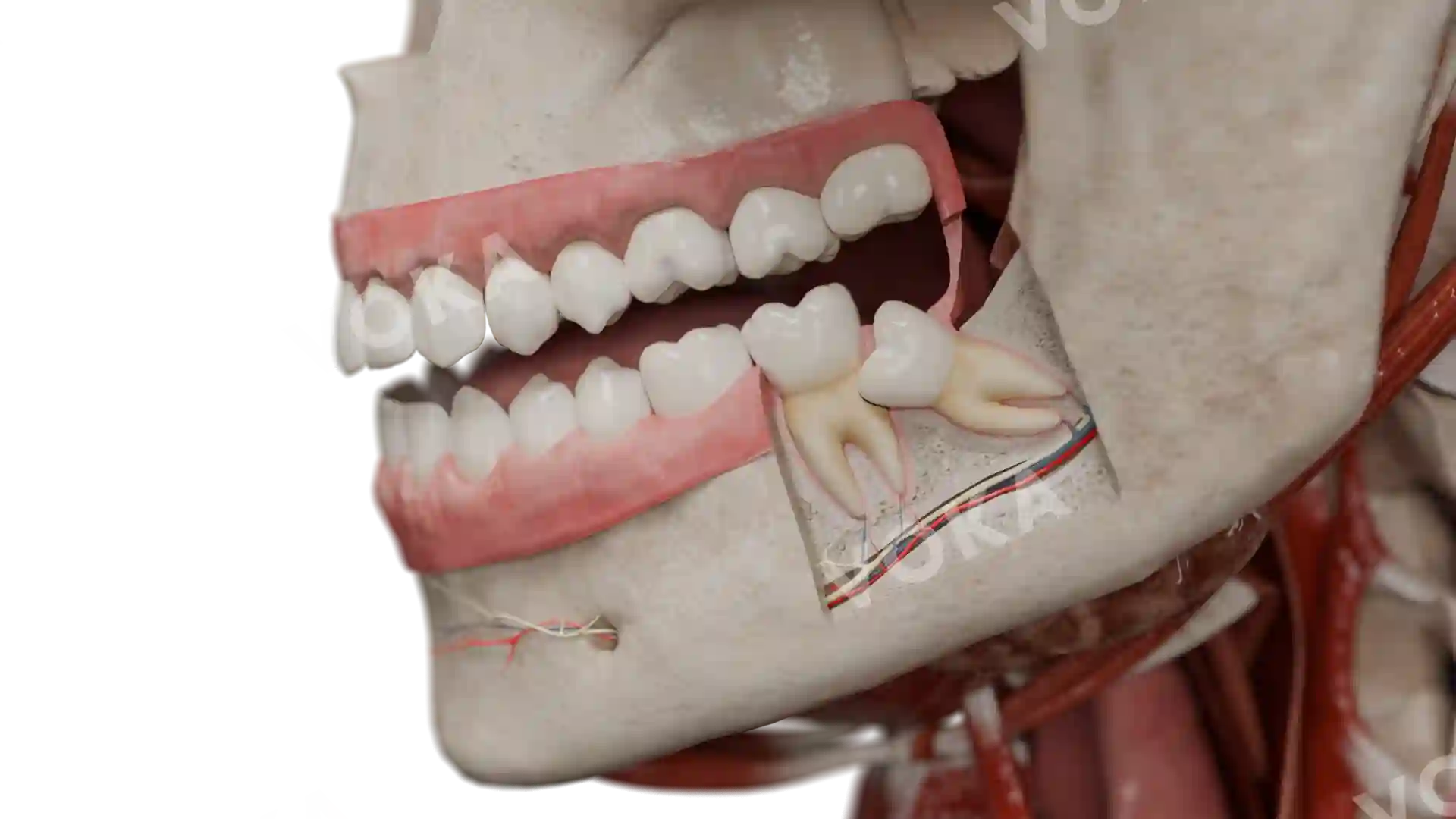
Anatomy of an acute apical abscess image - 30067

Dentistry

    License

    Commercial license grants the right to use an image for commercial purposes, such as advertising and marketing.

    Editorial license allows use for news, commentary, or non-commercial publications.

    Personal license grants permission for personal use, creating personal items like wall art.

    Print license authorizes the use of an image in physical print media, such as books, magazines, brochures, and posters.

    -Print

    Quantity

    Specify how many items you need. Bulk orders decrease the cost per unit.

    -More than 50

    Additional information

    Please be informed that when you click the Send message button VOKA Shop will process your personal data in accordance with our Privacy Policy.

    Anatomy of an acute apical abscess image

    Select license

    More information

    Details

    Background N/A
    Resolution 1920 x 1080 px
    Orientation Horizontal
    Format PNG
    File size 1.9 Mb
    Upload date June 10, 2025

    Item successfully added to the cart

    Link successfully copied to clipboard

    Thank you!

    Your message is sent!
    Our experts will contact you shortly. If you have any additional questions, please contact us at shop@voka.io