/Acute%20Otitis%20Media%20-%20Perforation%2C%202.webp)
/Acute%20Otitis%20Media%20-%20Perforation%2C%201.webp)


Description
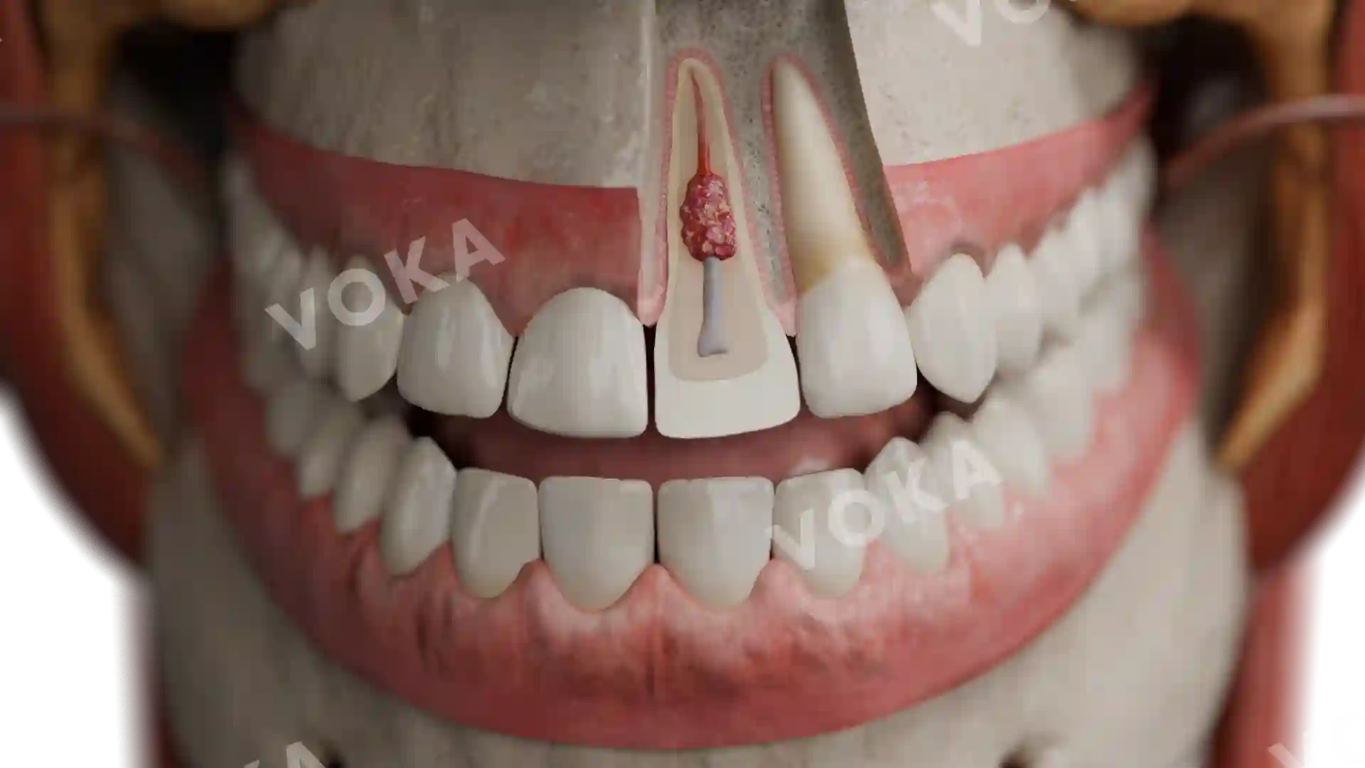
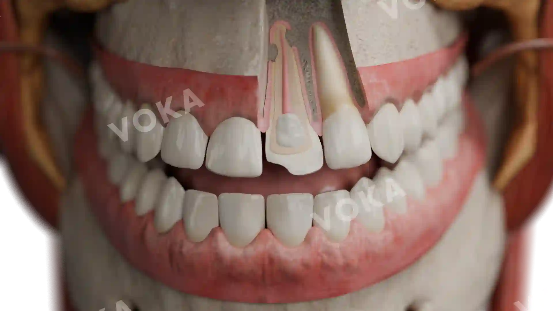
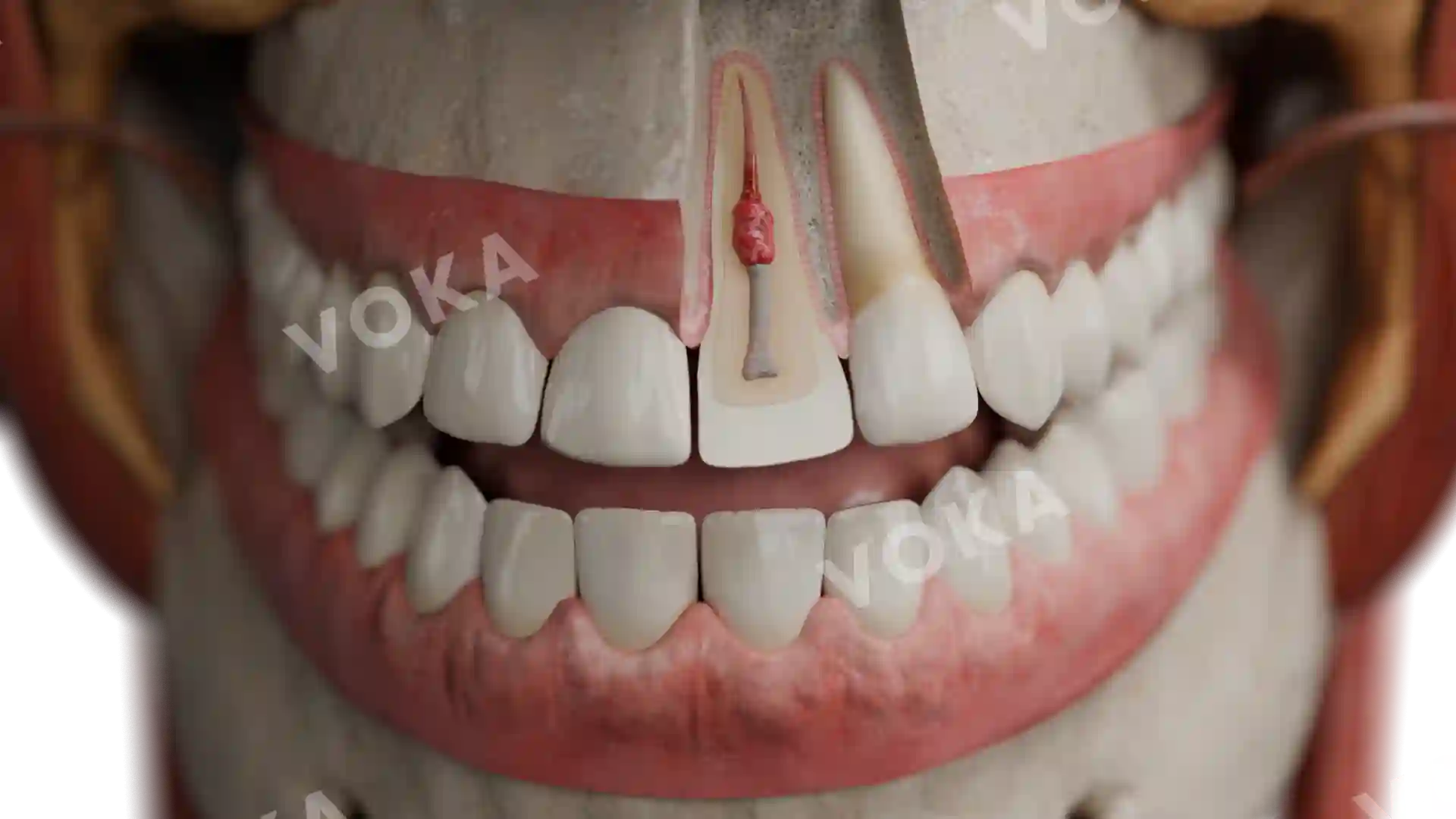
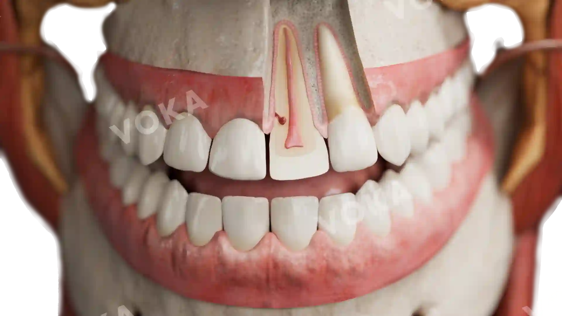
This sagittal section reveals the anatomy of an apical granuloma forming at the apex of a severely decayed lower molar. The tooth displays advanced structural damage from chronic caries, with infection having penetrated the pulp chamber and progressed through the root canal. At the tip of the root, the apical granuloma appears as a localized, soft tissue lesion consisting of granulation tissue, inflammatory cells, and fibrous tissue. This image provides a clear and instructive visualization of the pathological processes involved in periapical periodontitis and the formation of an apical granuloma.
Related items


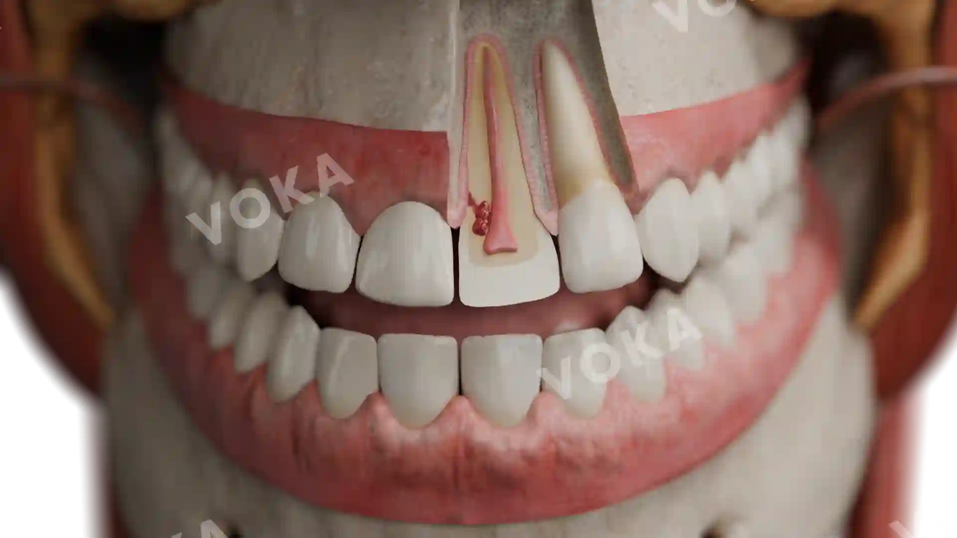
Anatomy of an apical granuloma image - 30063
Dentistry
Select license
More information
Details
Background
N/A
Resolution
1920 x 1080 px
Orientation
Horizontal
Format
PNG
File size
2.1 Mb
Upload date
June 10, 2025
Item successfully added to the cart