/Acute%20Otitis%20Media%20-%20Perforation%2C%202.webp)
/Acute%20Otitis%20Media%20-%20Perforation%2C%201.webp)


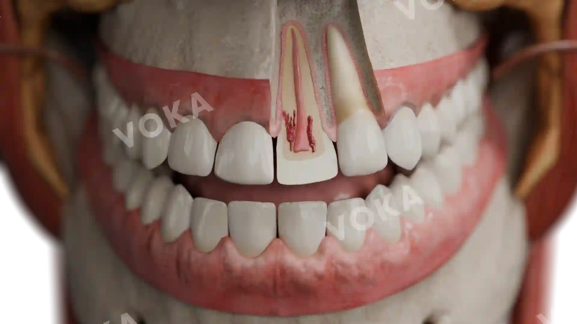
_(From_2_3%20of_the_length_of_the_dental_crown_to_the_gingiva).webp)
Description
This anatomical image illustrates advanced dental attrition, where the loss of hard tissue has progressed to approximately two-thirds of the crown height, approaching the gingival margin. The upper anterior teeth show significant wear with flattened, irregular incisal edges and exposed dentin, indicating chronic mechanical erosion from prolonged frictional contact—commonly seen in bruxism or age-related wear. Such attrition compromises both function and aesthetics, affecting speech, chewing, and vertical dimension of occlusion. This visual is especially valuable for clinicians and students in understanding severe cases of occlusal trauma, aiding in the diagnosis and treatment planning for full-mouth rehabilitation or protective occlusal therapy.
Related items
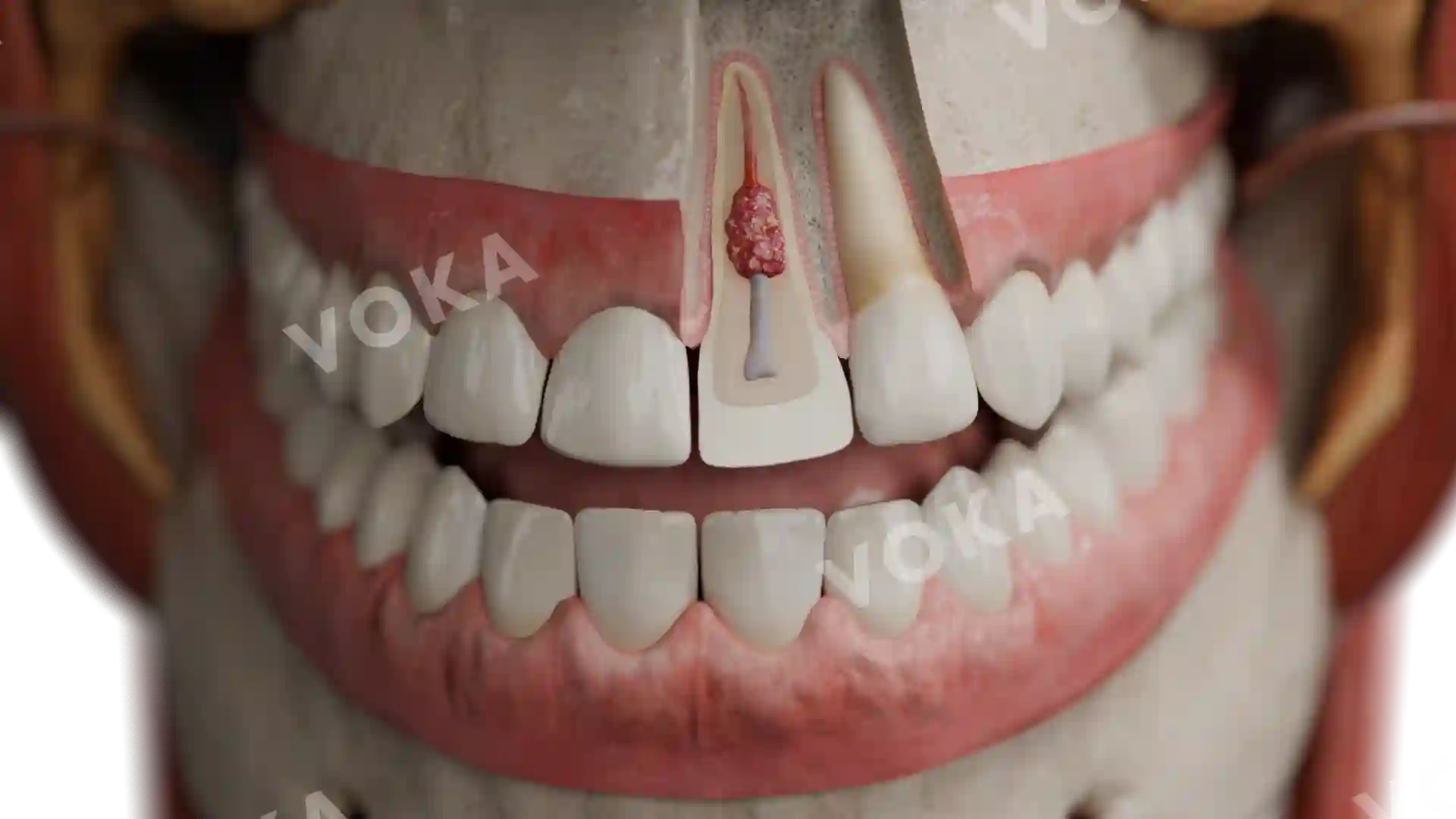


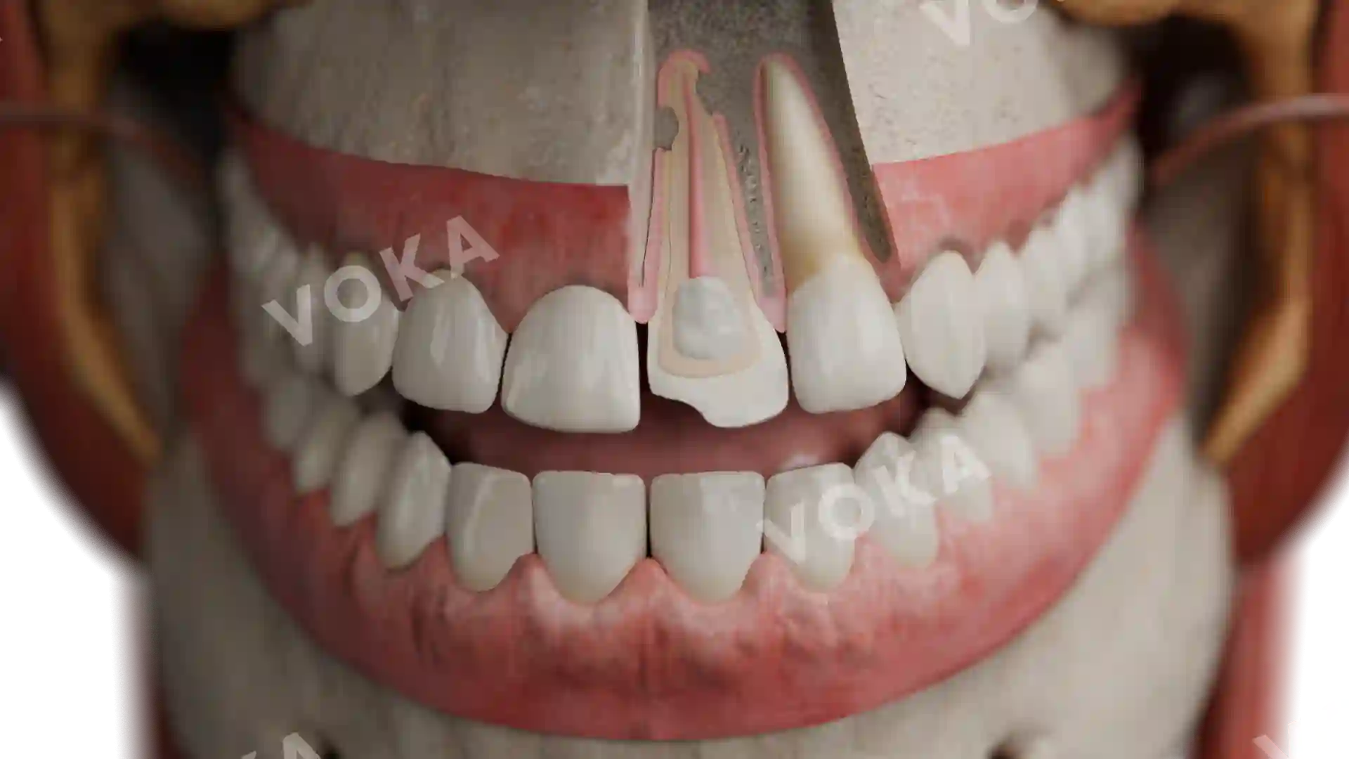
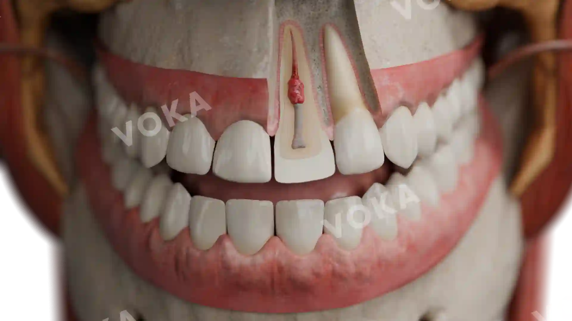
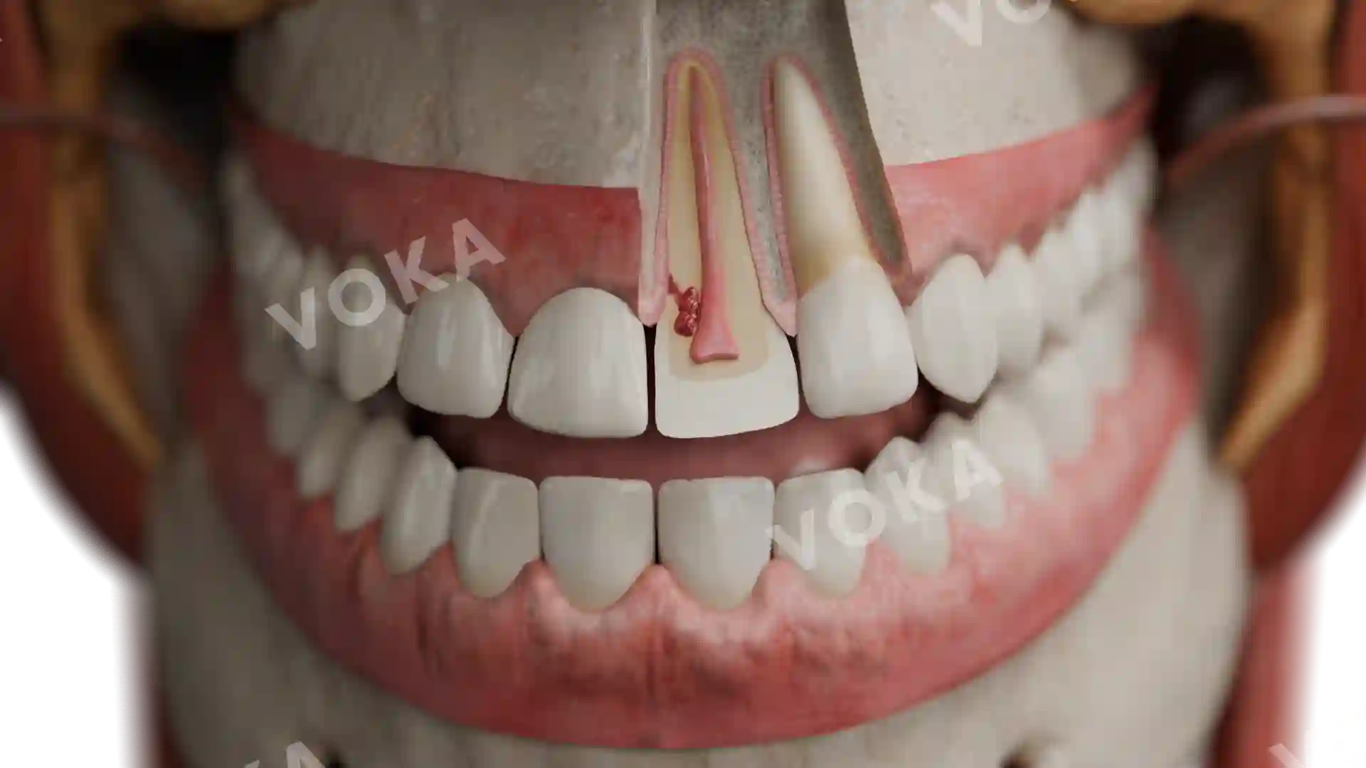
Dental attrition (depth of lesion from 2/3 of the length of the dental crown to the gingiva) image - 30050
Dentistry
Select license
More information
Details
Item successfully added to the cart