/Acute%20Otitis%20Media%20-%20Perforation%2C%202.webp)
/Acute%20Otitis%20Media%20-%20Perforation%2C%201.webp)


.webp)
Description
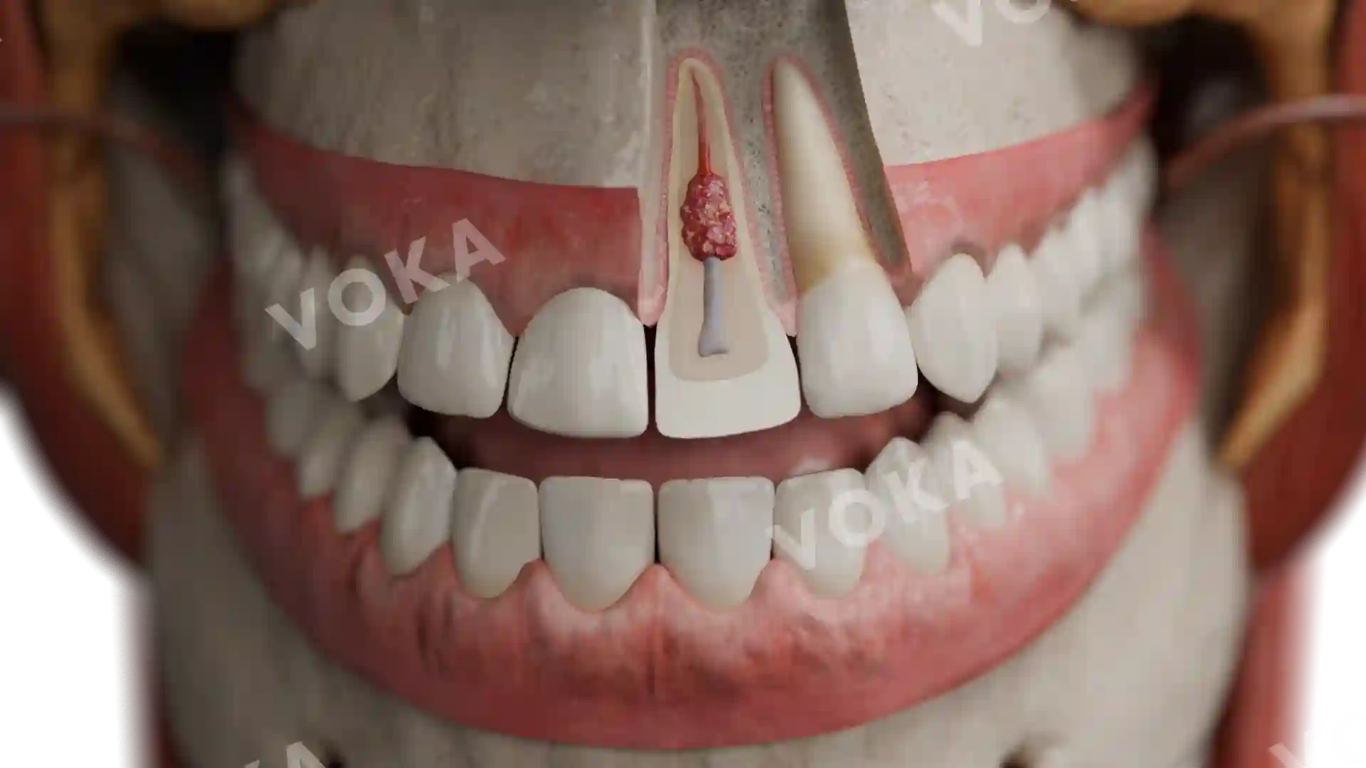
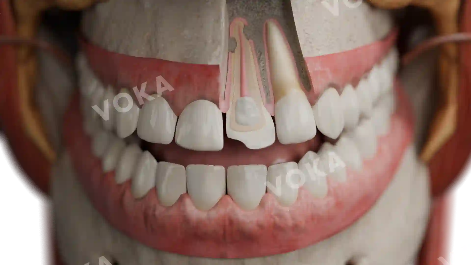
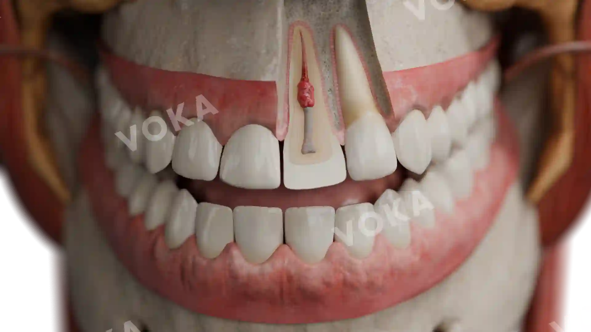
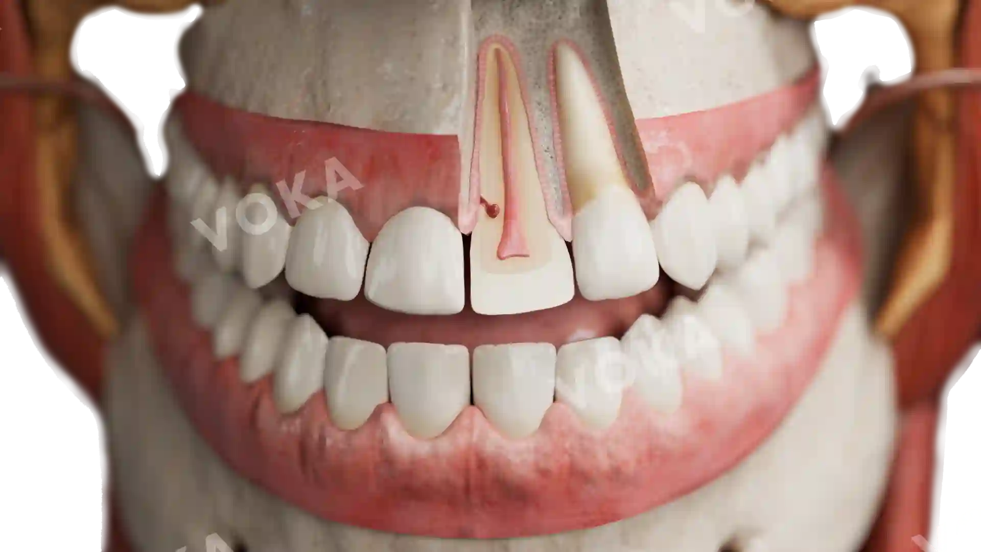
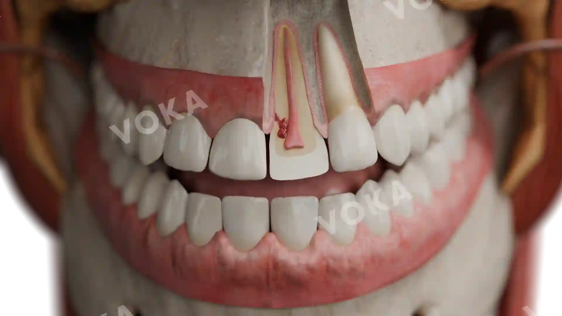
This anatomical image showcases a case of enamel hyperplasia on the buccal aspect of a posterior tooth. The enameloma appears as a small, rounded, enamel-covered projection near the root bifurcation, often resulting from developmental disturbances during the tooth formation stage. The detailed rendering clearly illustrates how this anomaly, though typically asymptomatic, can pose clinical challenges. This image serves as an excellent visual aid for dental students and clinicians to recognize such anatomical variations during diagnosis or treatment planning, particularly in cases involving periodontal or endodontic assessment.
Related items


Enamel hyperplasia (enameloma) image - 30053
Dentistry
Select license
More information
Details
Item successfully added to the cart