/Acute%20Otitis%20Media%20-%20Perforation%2C%202.webp)
/Acute%20Otitis%20Media%20-%20Perforation%2C%201.webp)


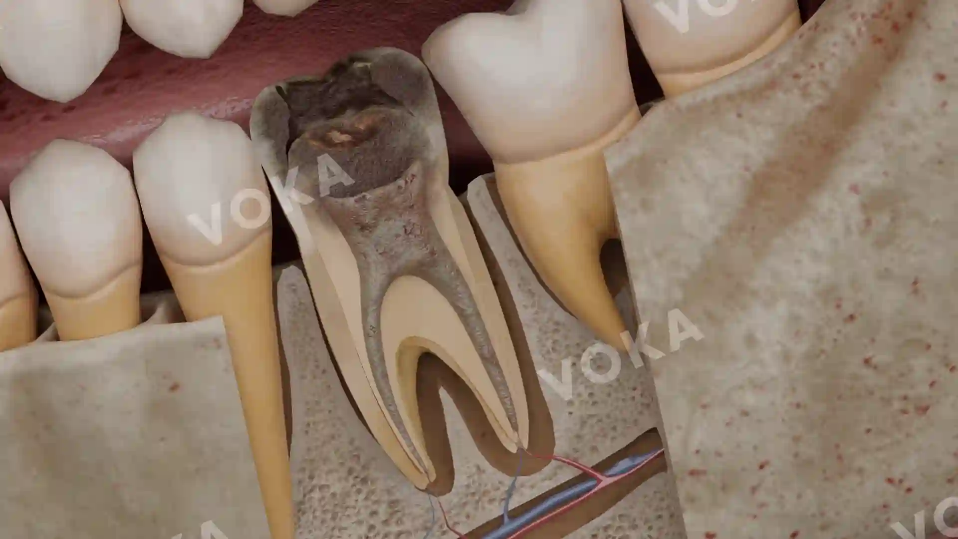
Description
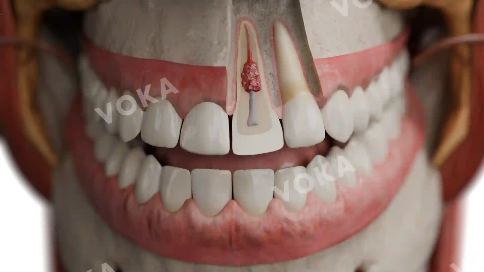
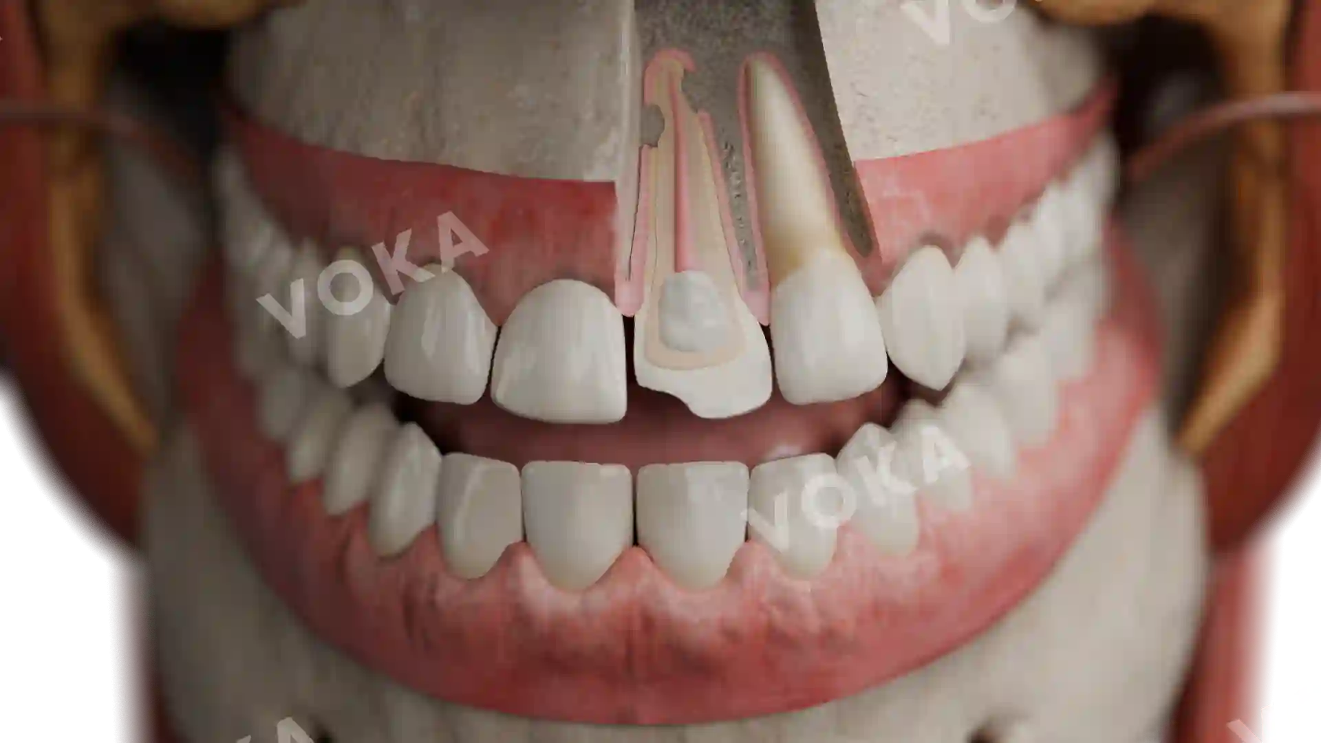
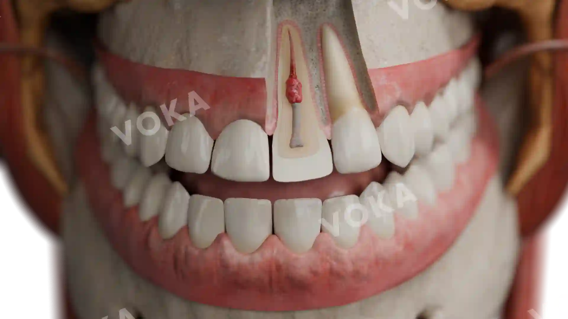
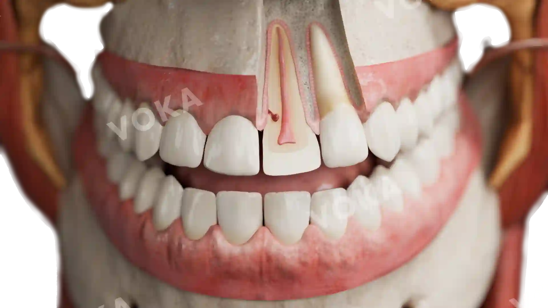
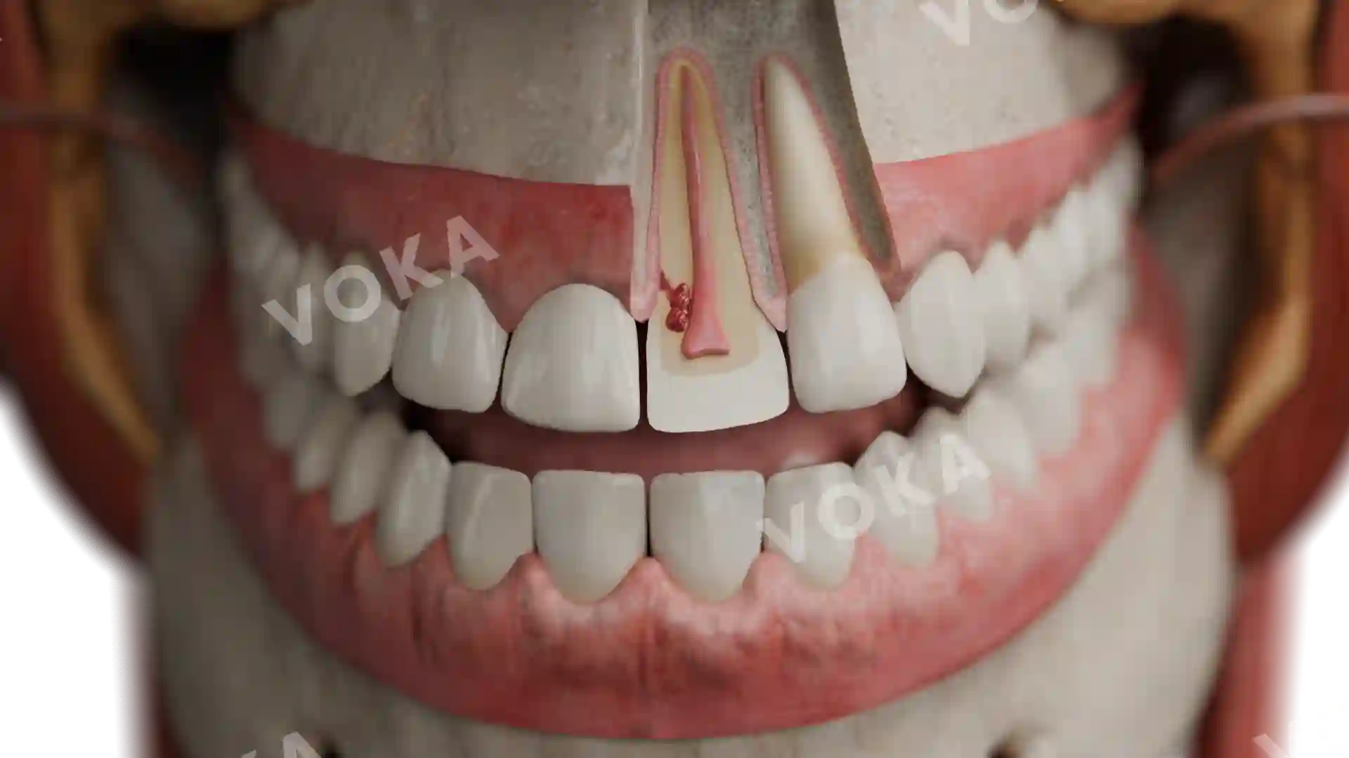
This detailed anatomical image provides a close-up view of tooth pulp necrosis, showcasing the internal structure of an affected tooth. The image highlights the severe degradation of the pulp tissue, visible as darkened, necrotic material within the pulp chamber and root canals. The vascular and nerve supply beneath the tooth illustrates how pulp necrosis can disrupt blood flow and nerve function, potentially leading to infection or abscess formation. This high-resolution image is an excellent resource for dental students, educators, and healthcare professionals, clearly visualizing the progression and impact of tooth pulp necrosis. Ideal for dental education, clinical presentations, and patient awareness, it simplifies the understanding of this common yet serious dental condition through precise, detailed visuals.
Related items


Necrosis of the tooth pulp image - 25026
Dentistry
Select license
More information
Details
Item successfully added to the cart