/Acute%20Otitis%20Media%20-%20Perforation%2C%202.webp)
/Acute%20Otitis%20Media%20-%20Perforation%2C%201.webp)


Description
This anatomical illustration depicts reversible pulpitis with visible signs of peripheral pulp edema and hyperemia. The sagittal section of the tooth reveals a deep carious lesion that has breached the enamel and reached close to the pulp chamber, triggering an early inflammatory response. The pulp tissue is noticeably reddened, indicating increased blood flow, and slightly swollen due to fluid accumulation. Despite the irritation, the overall structure of the pulp remains intact, suggesting that the condition is still potentially reversible with timely treatment. The surrounding periodontal tissues and bone show no pathological changes, underscoring the localized nature of the inflammation at this stage.
Related items
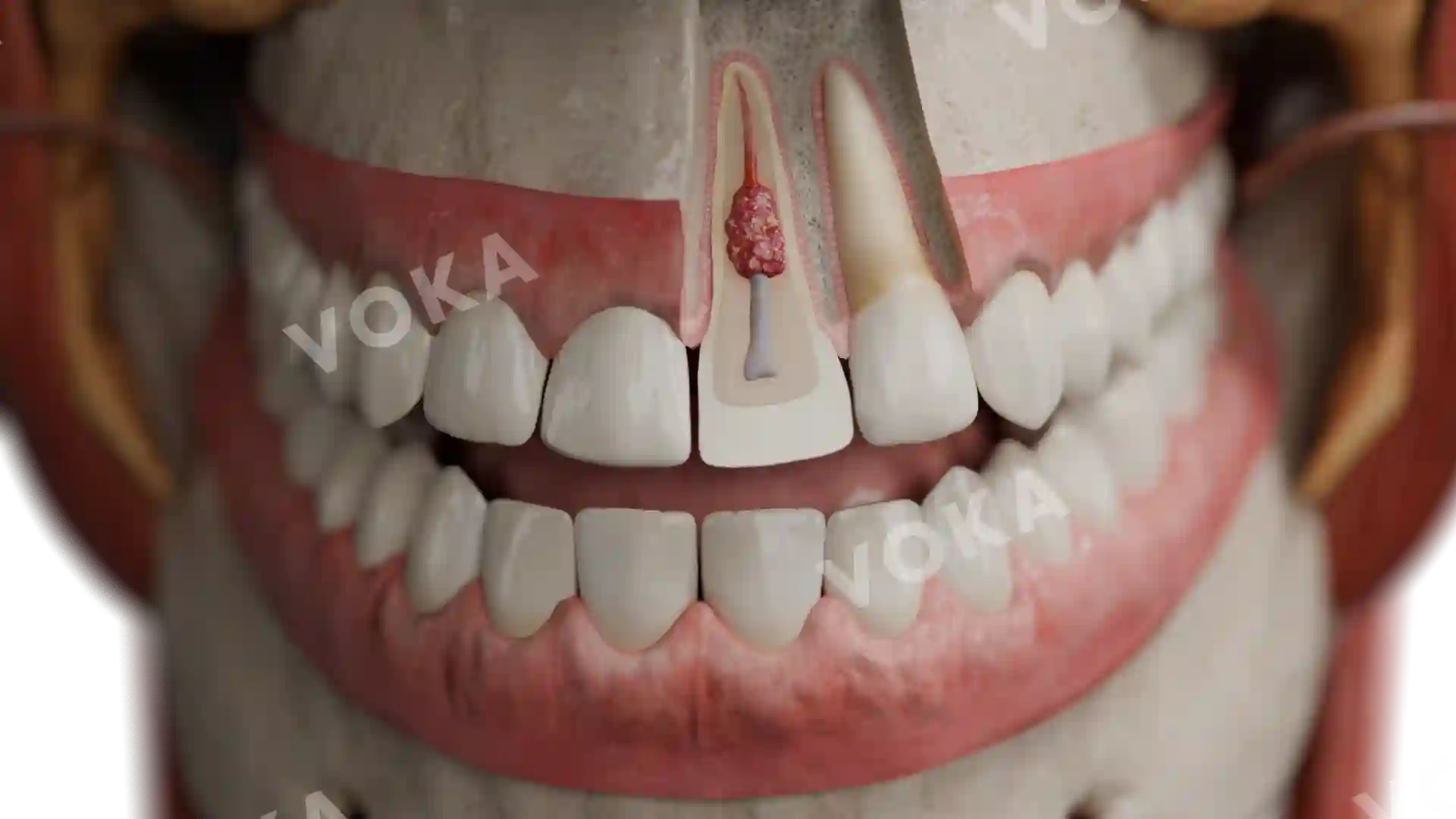


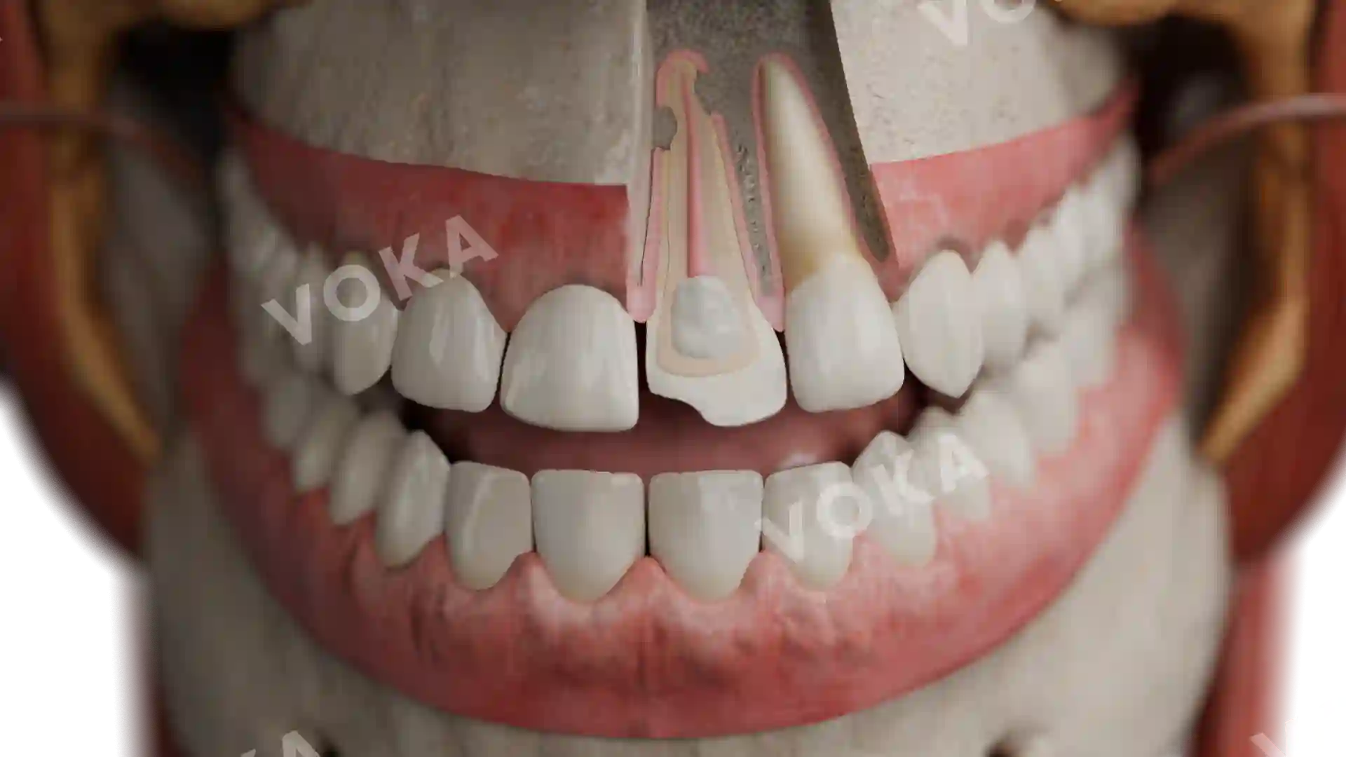
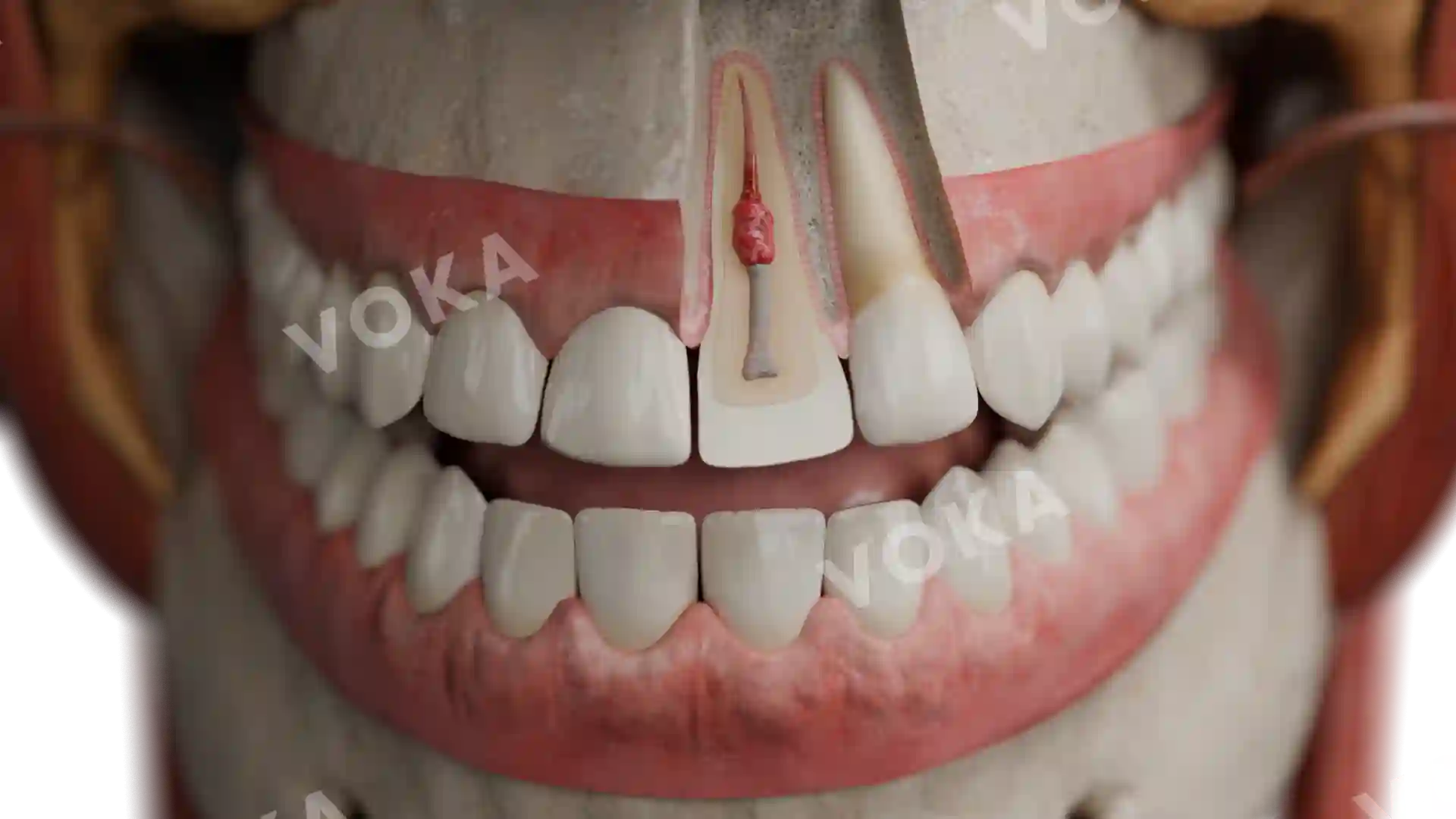
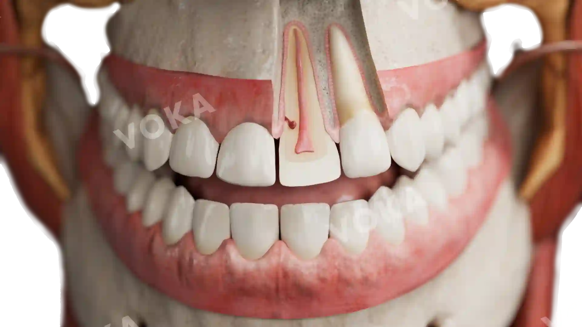
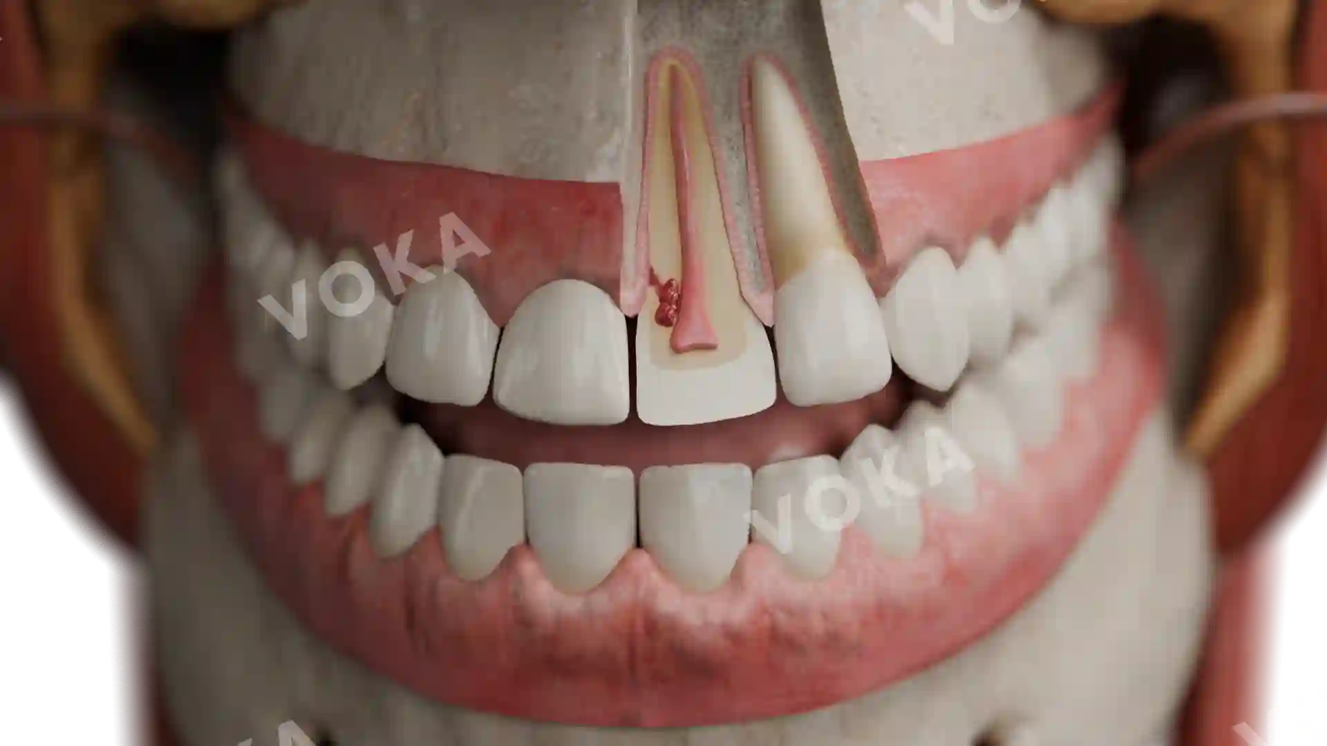
Peripheral pulp edema and hyperemia in reversible pulpitis image - 30070
Dentistry
Select license
More information
Details
Item successfully added to the cart