/Acute%20Otitis%20Media%20-%20Perforation%2C%202.webp)
/Acute%20Otitis%20Media%20-%20Perforation%2C%201.webp)


Description
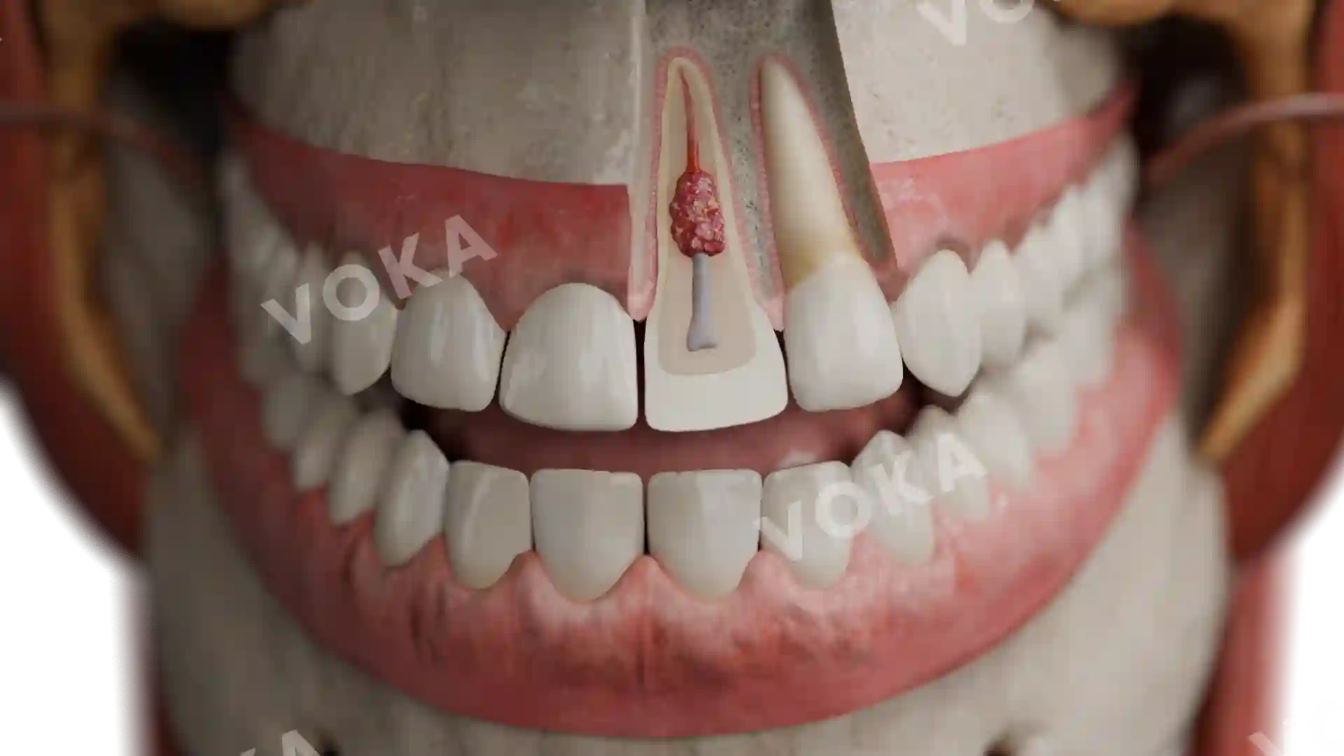
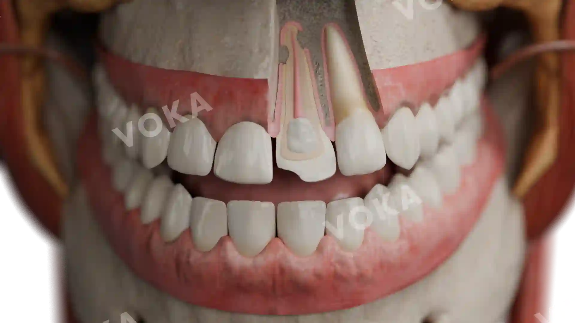
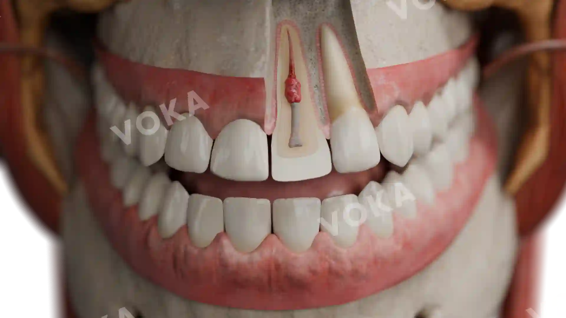
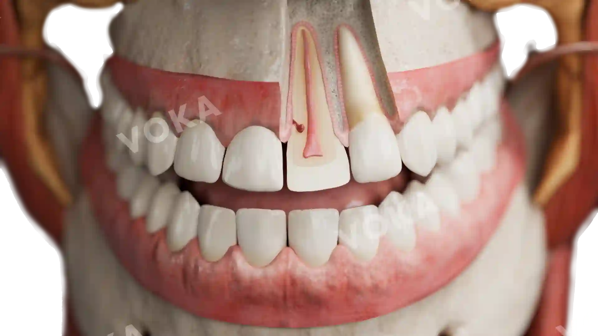
This high-resolution image captures the presence of secondary caries around a restoration in a maxillary molar. The darkened margins surrounding the filling indicate areas of recurrent decay, where bacterial infiltration has compromised the interface between the dental restoration and the natural tooth structure. This condition typically arises when plaque accumulates at the edges of a filling, leading to demineralization and subsequent carious lesions beneath or adjacent to the restoration. Ideal for educational and diagnostic purposes, this visualization offers a realistic and detailed representation of how secondary caries develop in clinical practice, thereby reinforcing the importance of ongoing maintenance for restored teeth.
Related items


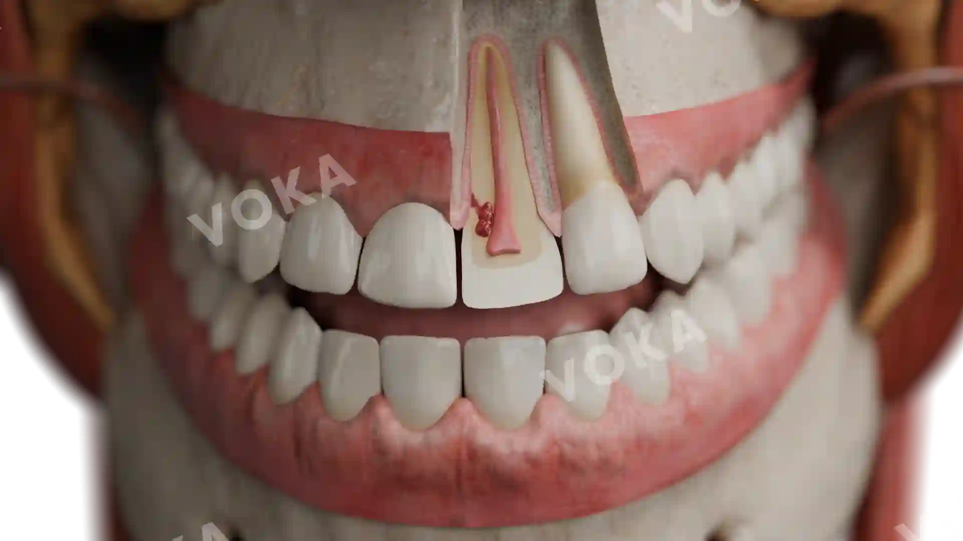
Secondary caries of maxillary molar image - 30040
Dentistry
Select license
More information
Details
Item successfully added to the cart