/Acute%20Otitis%20Media%20-%20Perforation%2C%202.webp)
/Acute%20Otitis%20Media%20-%20Perforation%2C%201.webp)


Description
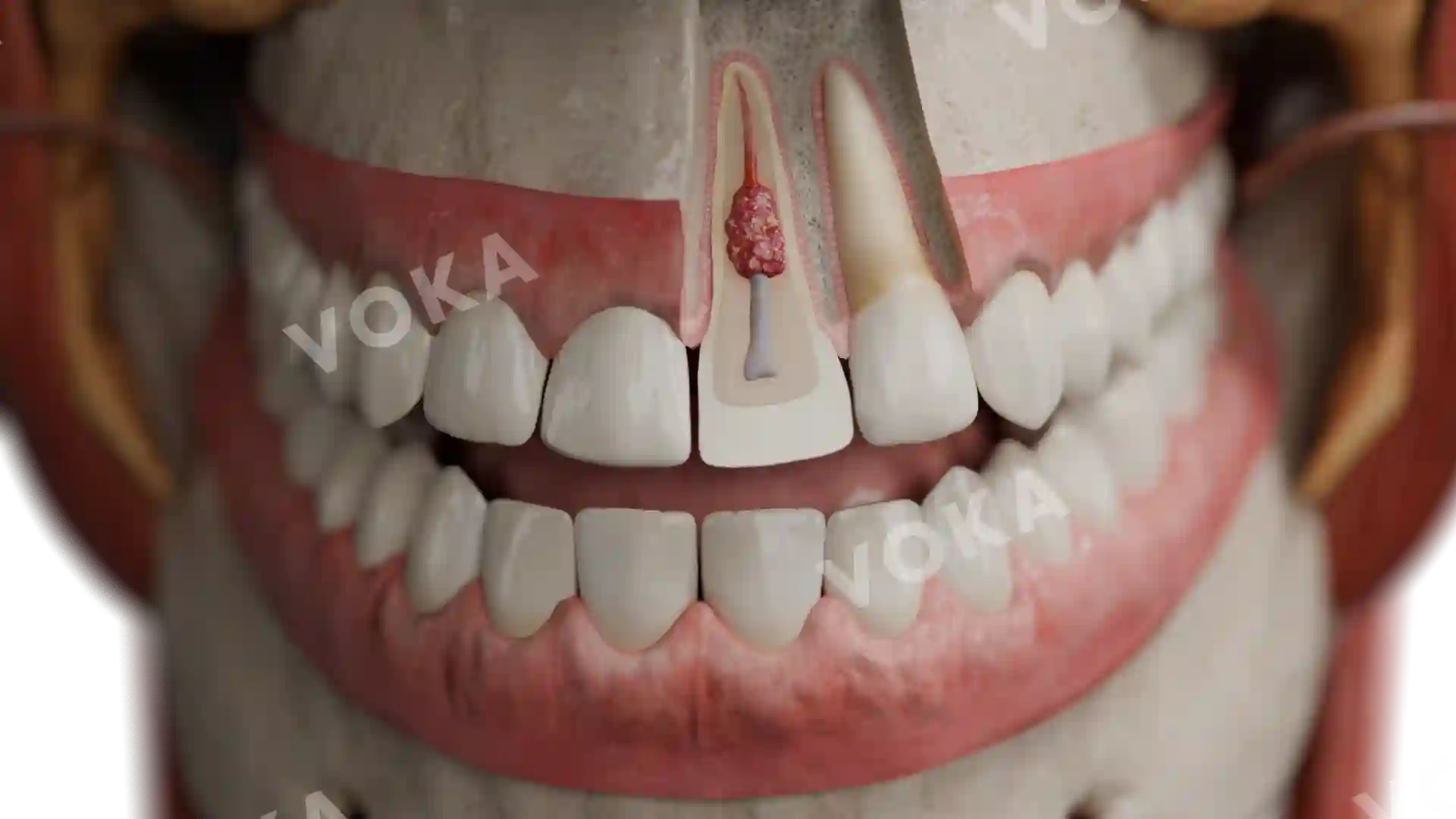
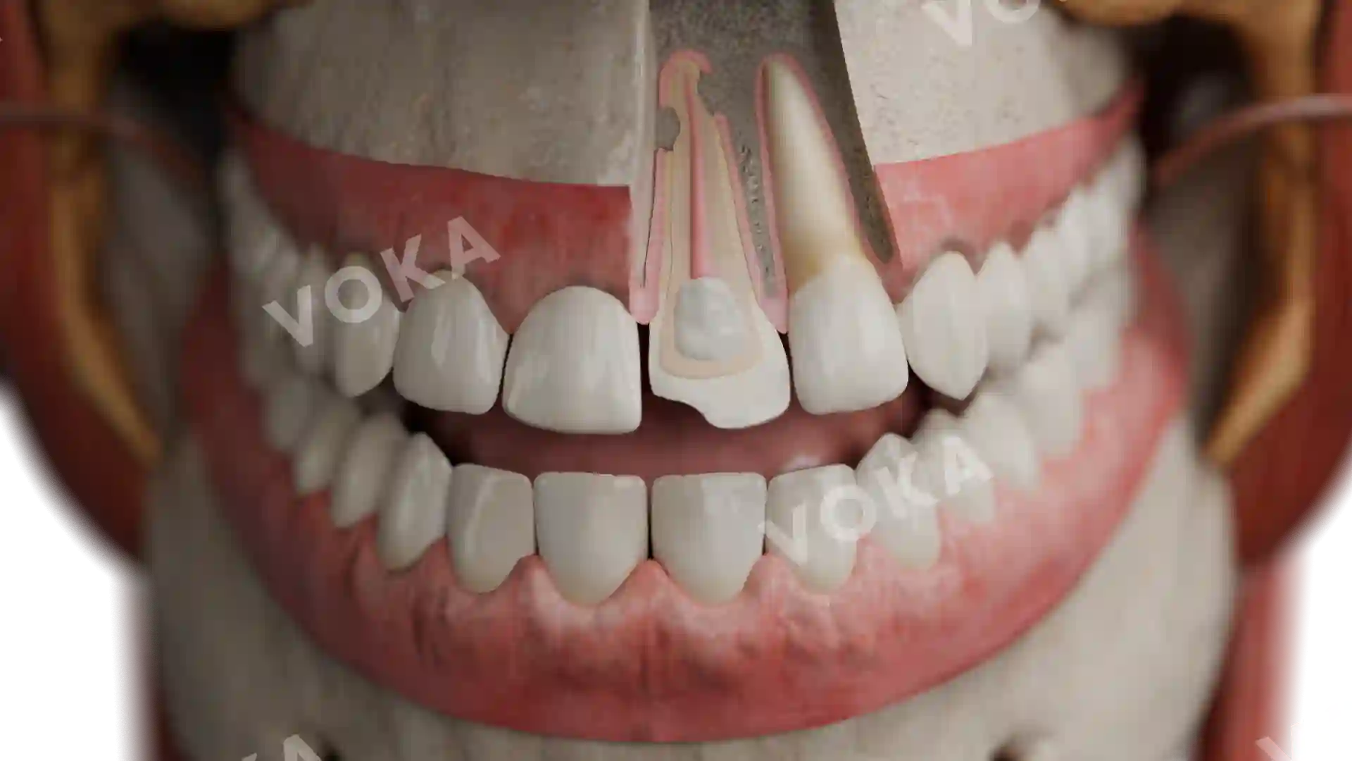
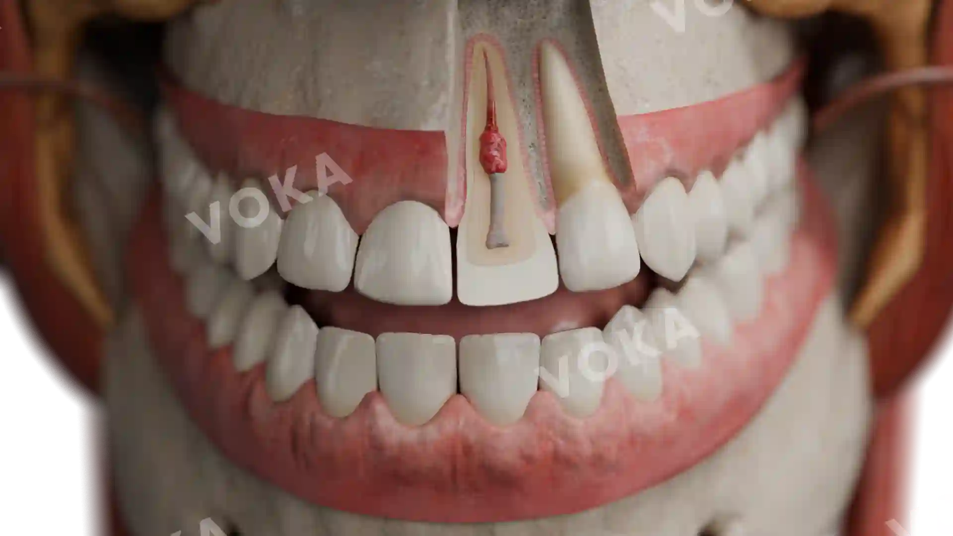
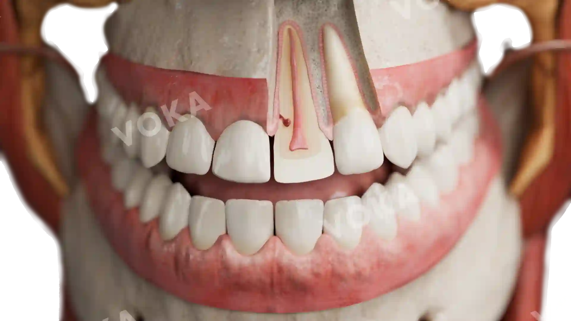
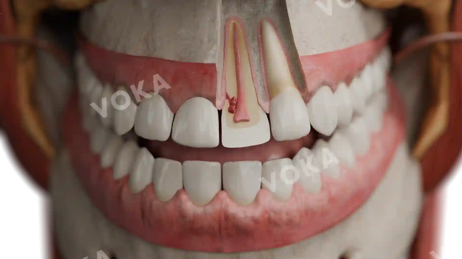
This detailed anatomical image provides a frontal view of systemic enamel hypoplasia, highlighting the characteristic linear defects across the teeth. The enamel exhibits horizontal grooves and pits, signs of disrupted enamel formation during tooth development. The enamel appears thin, with areas of discoloration and uneven surfaces, exposing dentin beneath in some regions. Surrounding anatomical structures, including facial muscles, nerves, blood vessels, and lymph nodes, are clearly visible, providing context for the oral cavity’s complex anatomy. Ideal for dental pathology courses, clinical presentations, and educational materials, it enhances the study of developmental enamel defects and their systemic implications.
Related items


Systemic enamel hypoplasia image - 25027
Dentistry
Select license
More information
Details
Item successfully added to the cart