/Acute%20Otitis%20Media%20-%20Perforation%2C%202.webp)
/Acute%20Otitis%20Media%20-%20Perforation%2C%201.webp)


Description
This detailed anatomical illustration depicts duodenal ulcers, visualized from a cross-sectional view of the duodenum. The image clearly shows multiple inflamed and eroded areas on the mucosal lining, characterized by localized redness, swelling, and crater-like lesions. The texture of the surrounding healthy tissue contrasts starkly with the ulcerated zones, emphasizing the pathological changes. This visual effectively highlights the classic appearance of peptic ulceration within the first portion of the small intestine. It is ideal for educational purposes in explaining mucosal breakdown caused by gastric acid exposure.
Related items


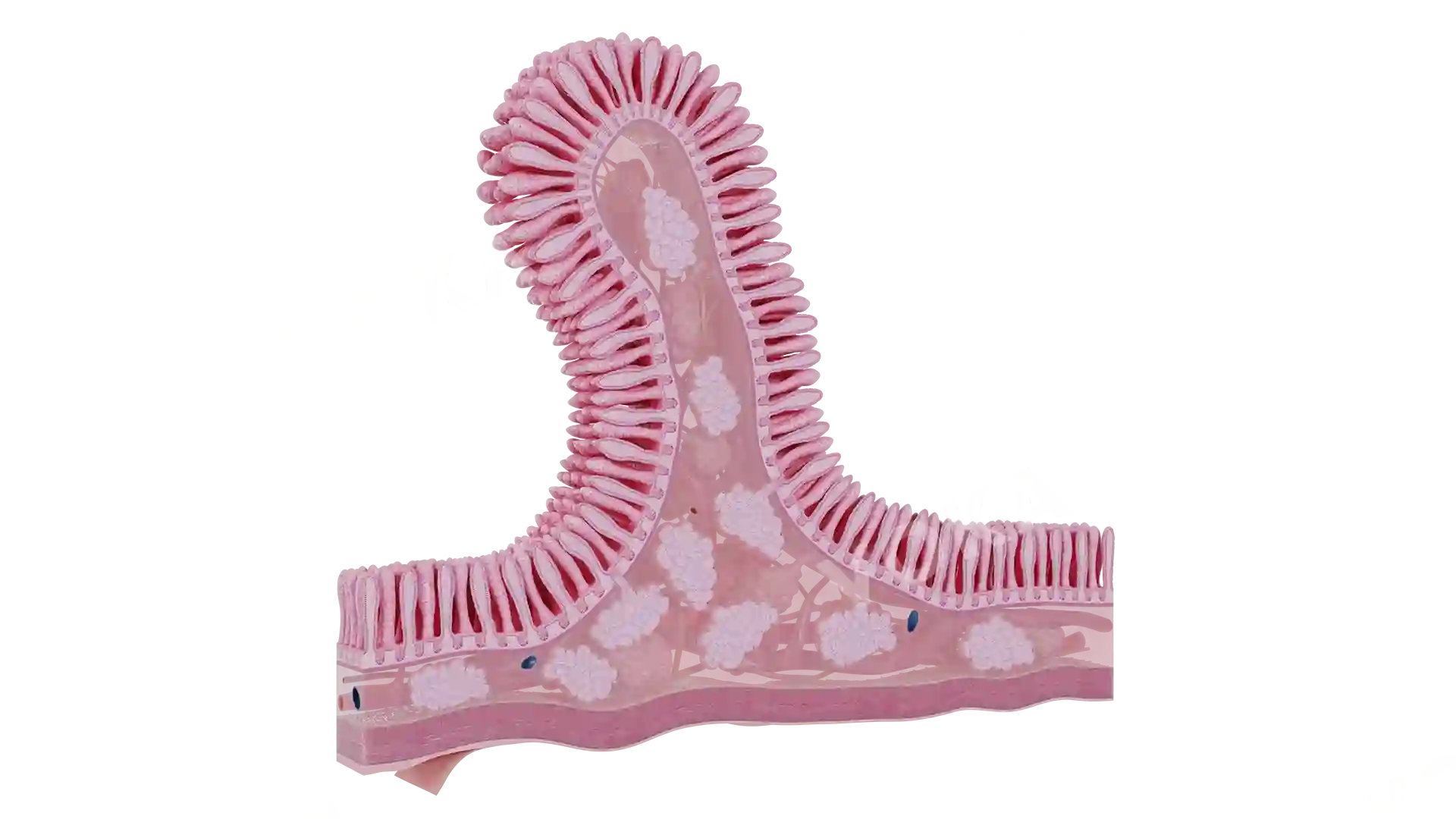


Duodenal Ulcers image - 30072
Gastroenterology
Select license
More information
Details
Item successfully added to the cart