/Acute%20Otitis%20Media%20-%20Perforation%2C%202.webp)
/Acute%20Otitis%20Media%20-%20Perforation%2C%201.webp)


Description
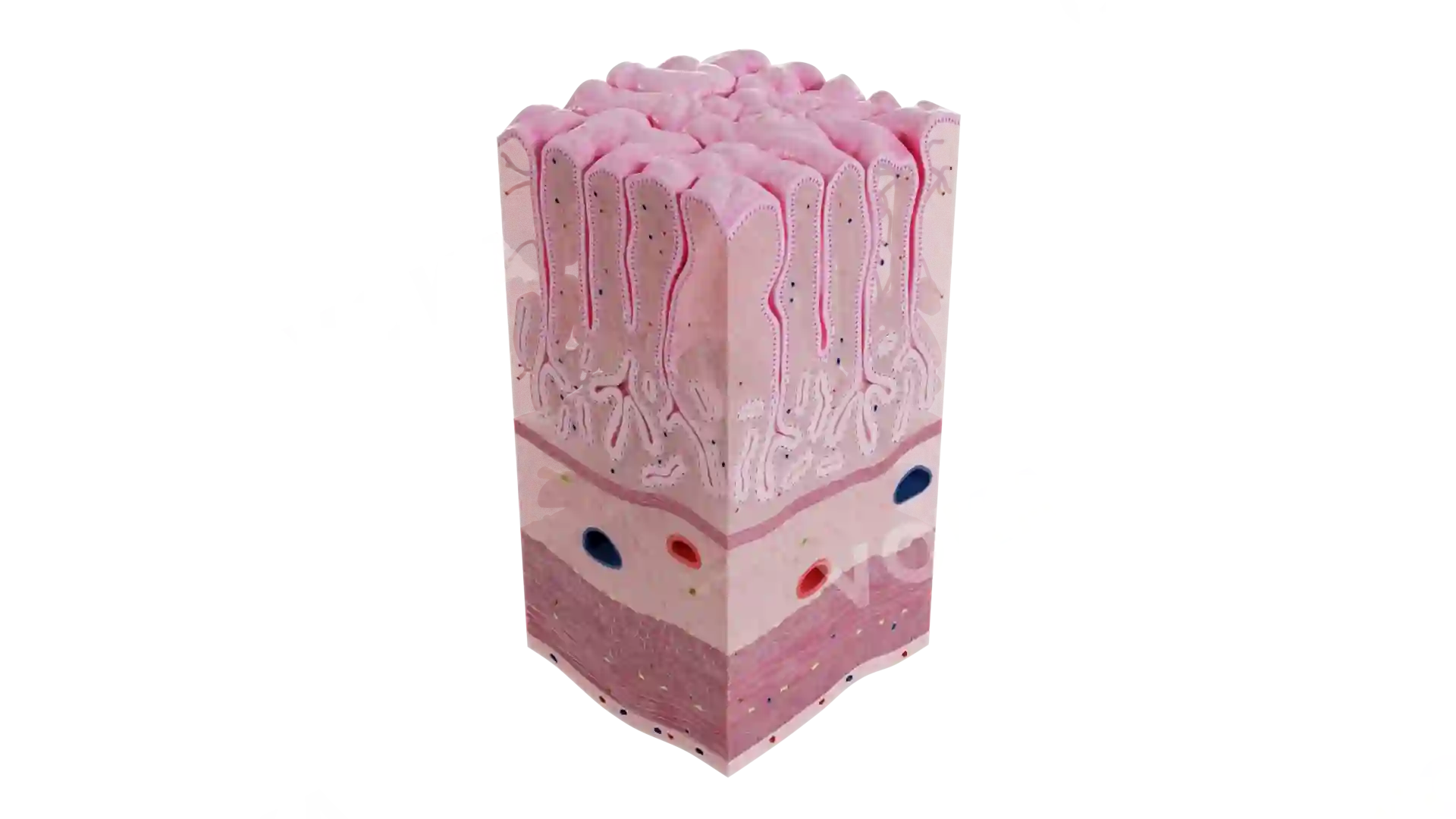
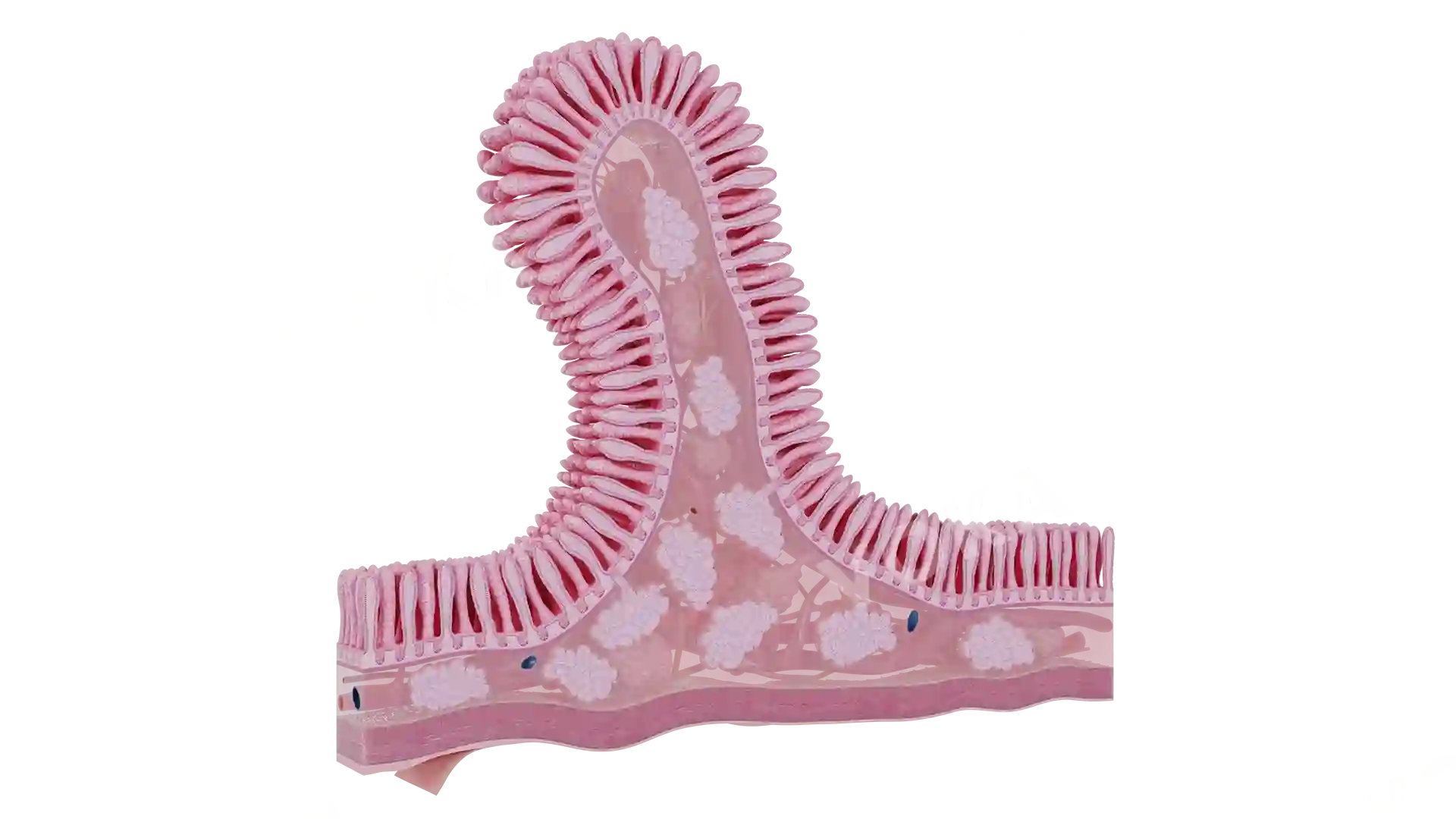
This anatomical illustration vividly captures erosive esophagitis, focusing on the lower portion of the esophagus and its transition into the stomach. The inner lining of the esophagus appears inflamed, with visible areas of mucosal erosion and redness indicative of acid-induced injury. The damaged surface contrasts with the relatively smooth gastric mucosa, emphasizing the pathological transition at the gastroesophageal junction. This detailed cross-sectional view is ideal for understanding the visual characteristics of esophageal mucosal damage often seen in chronic acid reflux conditions.
Related items


Erosive Esophagitis image - 30073
Gastroenterology
Select license
More information
Details
Background
Transparent
Resolution
1920 x 1080 px
Orientation
Horizontal
Format
PNG
File size
1.6 Mb
Upload date
June 10, 2025
Item successfully added to the cart