/Acute%20Otitis%20Media%20-%20Perforation%2C%202.webp)
/Acute%20Otitis%20Media%20-%20Perforation%2C%201.webp)


Description
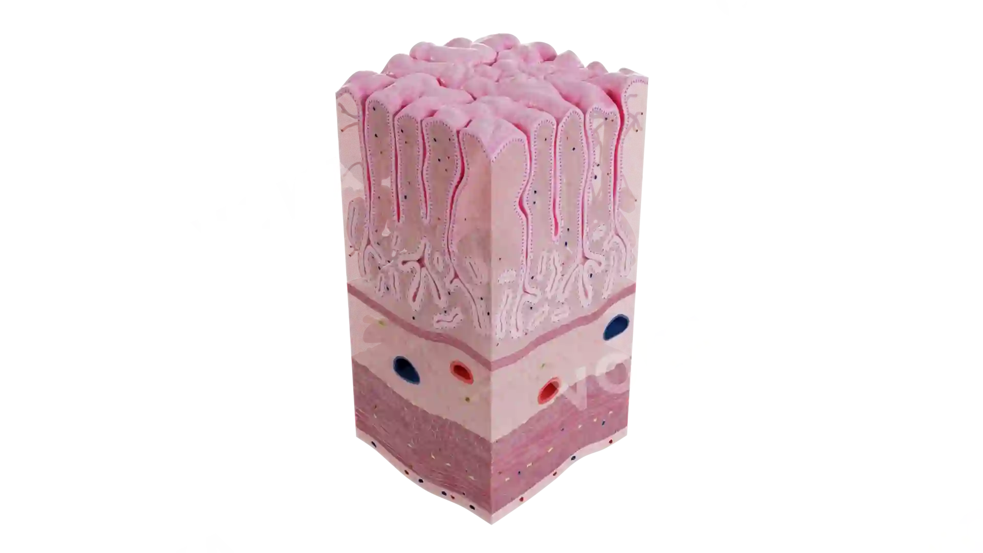
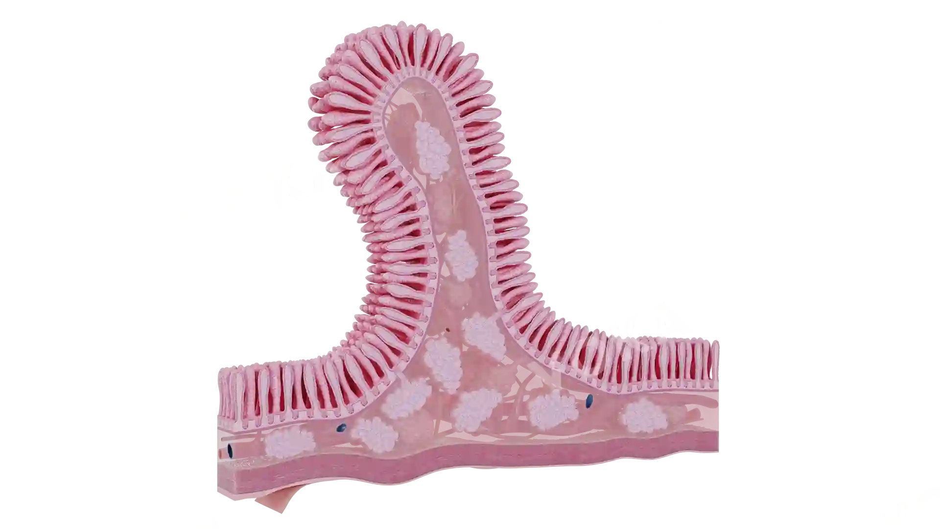
This anatomical illustration depicts Grade A erosive esophagitis, characterized by minimal mucosal damage at the gastroesophageal junction. The image shows a small, sharply demarcated erosion confined to a single mucosal fold without bridging across folds. The esophageal lining appears reddened and inflamed, with a visible localized break that highlights the initial stage of mucosal injury caused by acid reflux. The clarity and anatomical accuracy make this illustration particularly useful for medical education on the early detection and diagnosis of erosive esophagitis.
Related items


Grade A Erosive Esophagitis image - 30075
Gastroenterology
Select license
More information
Details
Background
Transparent
Resolution
1920 x 1080 px
Orientation
Horizontal
Format
PNG
File size
1.5 Mb
Upload date
June 10, 2025
Item successfully added to the cart