/Acute%20Otitis%20Media%20-%20Perforation%2C%202.webp)
/Acute%20Otitis%20Media%20-%20Perforation%2C%201.webp)


Description
This anatomical illustration represents Grade B erosive esophagitis, highlighting visible mucosal breaks at the gastroesophageal junction. The erosions here span across multiple mucosal folds, while still measuring under 5 mm in length. The image clearly shows inflamed and reddened tissue, with disrupted surface continuity suggestive of acid-induced damage. This detailed cross-section offers an excellent visual reference for understanding moderate esophageal involvement in reflux disease, aiding in clinical recognition and educational discussion.
Related items


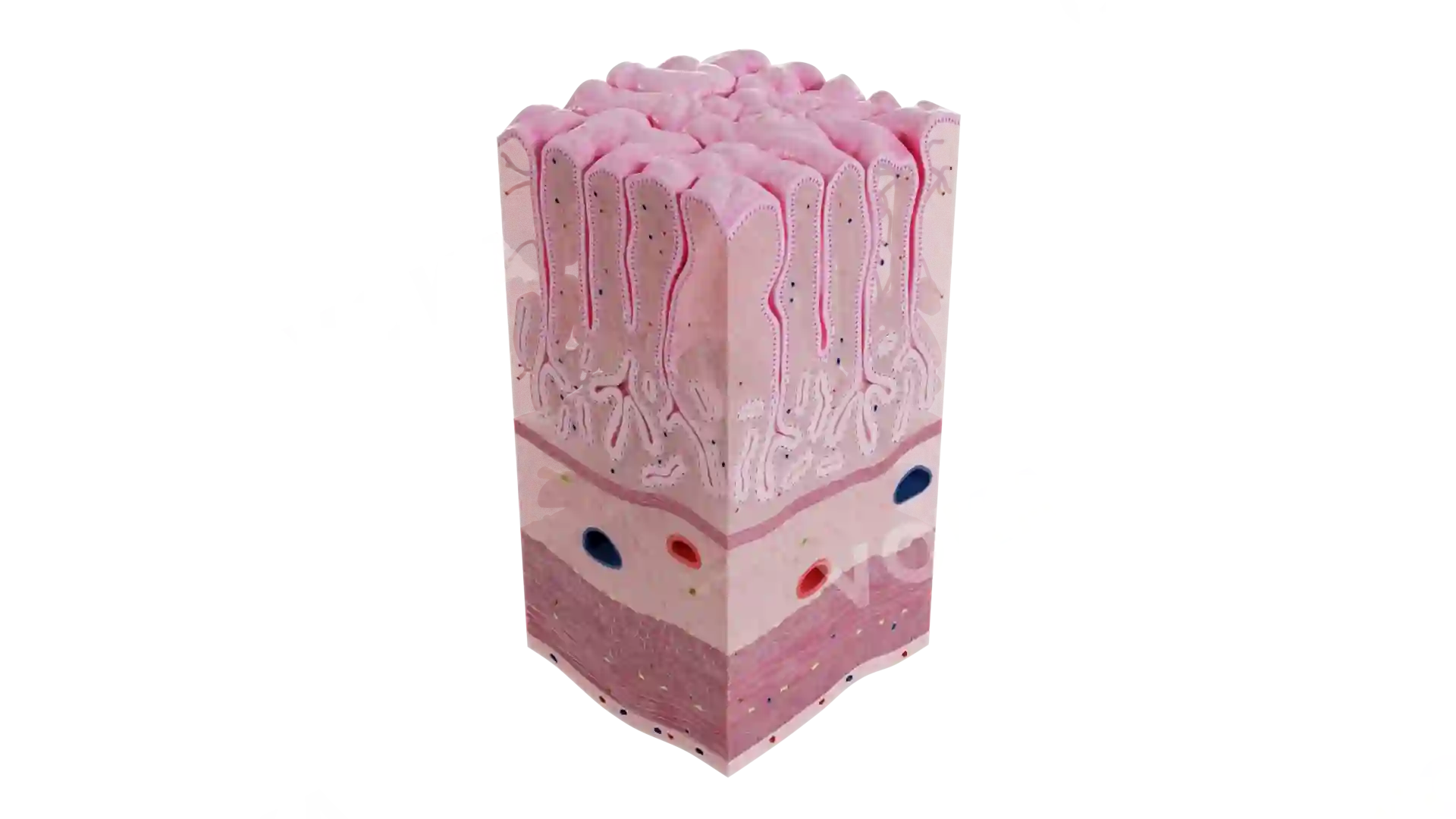
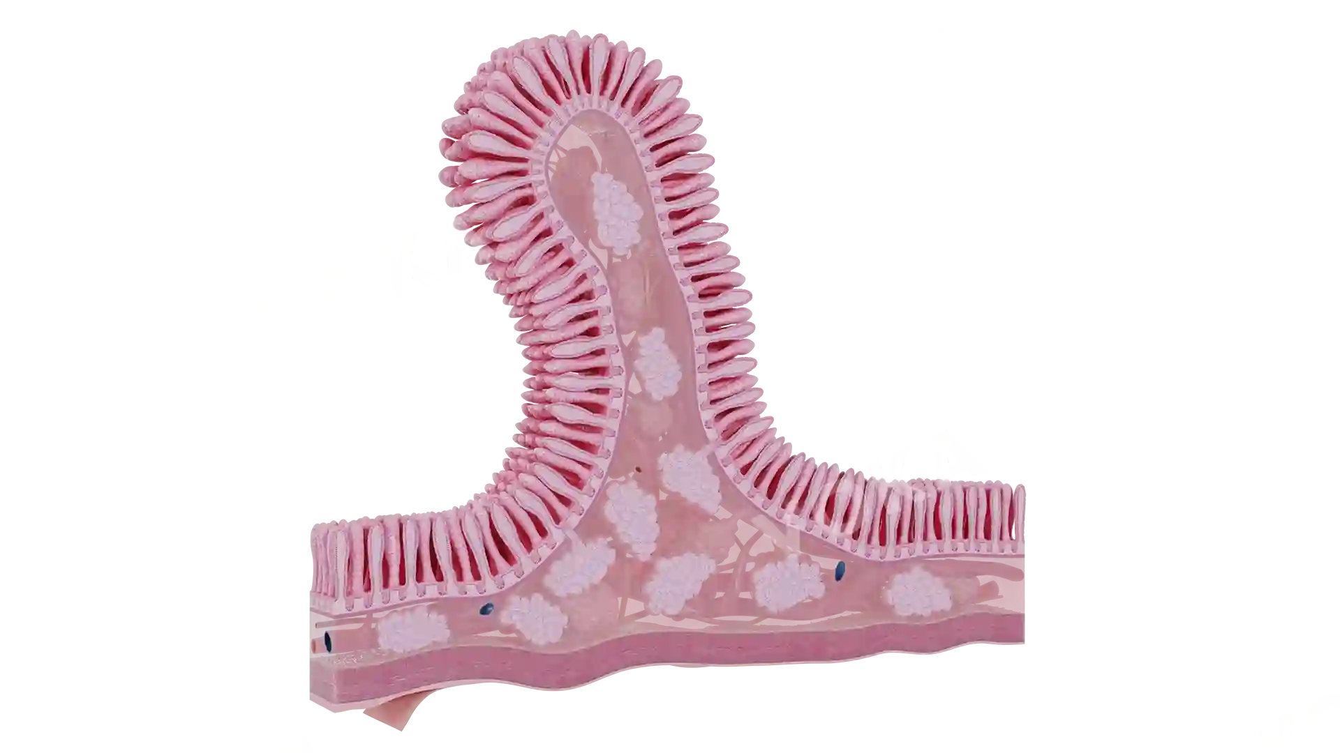


Grade B Erosive Esophagitis image - 30076
Gastroenterology
Select license
More information
Details
Background
Transparent
Resolution
1920 x 1080 px
Orientation
Horizontal
Format
PNG
File size
1.6 Mb
Upload date
June 10, 2025
Item successfully added to the cart