/Acute%20Otitis%20Media%20-%20Perforation%2C%202.webp)
/Acute%20Otitis%20Media%20-%20Perforation%2C%201.webp)


.webp)
Description
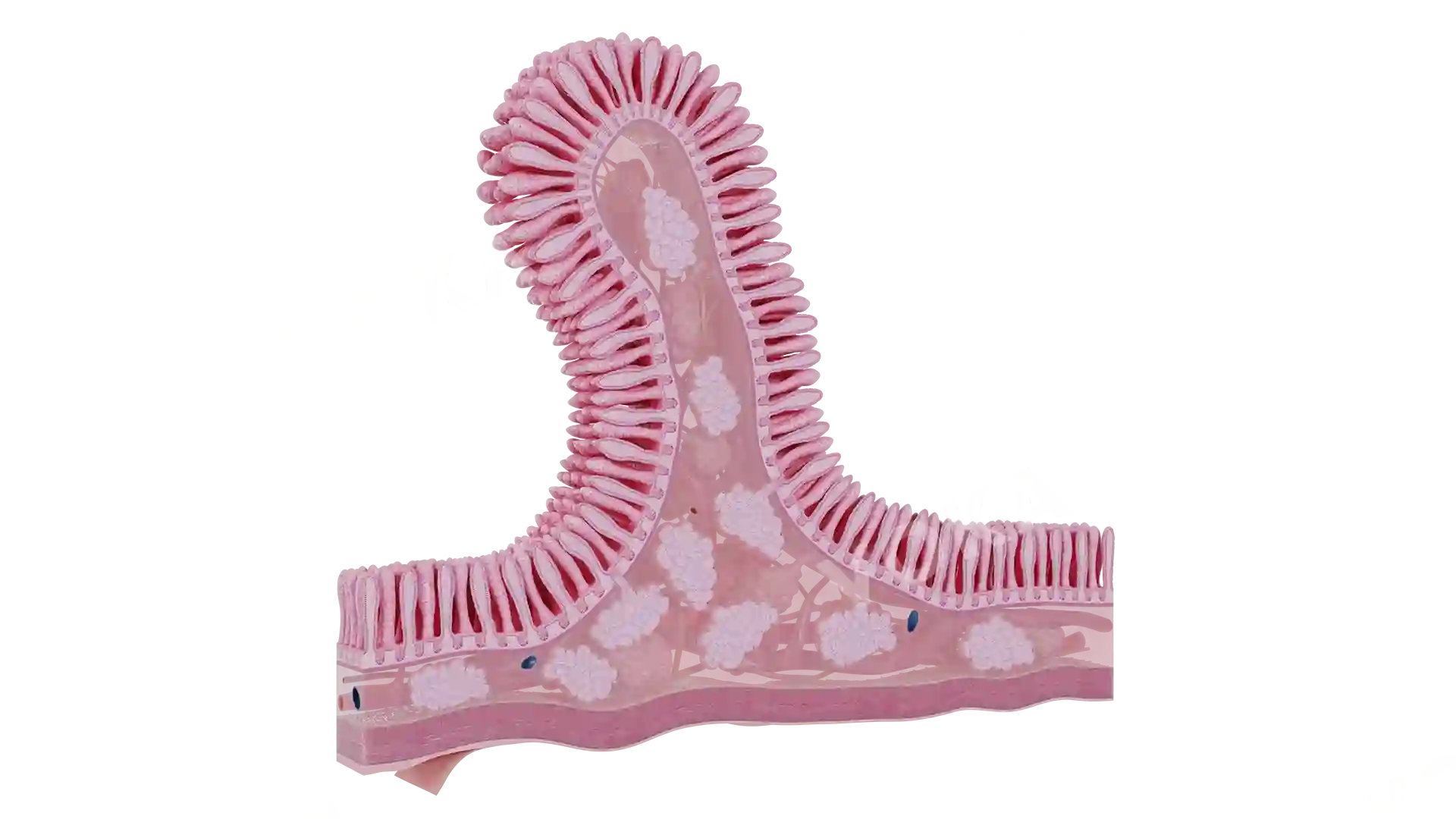
This detailed close-up image of the wall of the large intestine offers an in-depth view of its structure, highlighting the various layers that contribute to its function. The mucosal layer, featuring distinctive folds and villi, is clearly visible, aiding in the absorption of water and electrolytes. The muscular layer, which plays a key role in peristalsis, can also be seen, demonstrating how it contracts to move waste through the colon. The outer layer of connective tissue helps to support and protect the intestine. This image is an excellent resource for studying gastrointestinal anatomy, digestive processes, and pathologies like colitis or diverticulosis.
Related items


Wall of the large intestine (close up) image - 25240
Gastroenterology
Select license
More information
Details
Item successfully added to the cart