/Acute%20Otitis%20Media%20-%20Perforation%2C%202.webp)
/Acute%20Otitis%20Media%20-%20Perforation%2C%201.webp)


Description
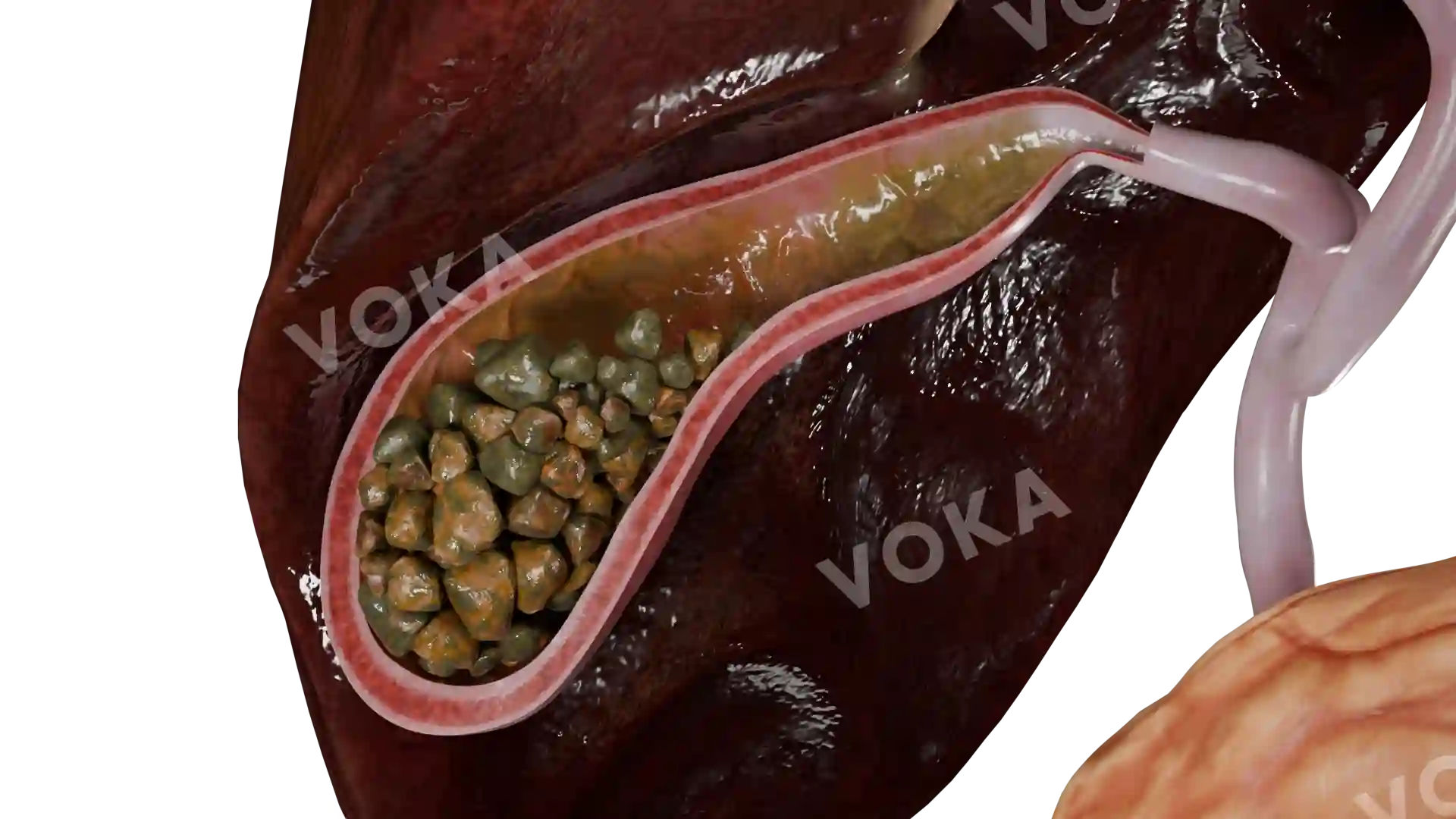
Investigate mixed liver cirrhosis using this high-resolution 3D model, demonstrating a combination of small and large nodules interspersed with fibrotic tissue throughout the hepatic parenchyma. The visualization emphasizes heterogeneity in nodule size, distribution, and fibrous septa, showing distortion of normal lobular and vascular anatomy. The model provides a clear perspective on the anatomical consequences of chronic liver injury, highlighting interactions between regenerative nodules, connective tissue, and blood vessels. It is valuable for studying morphological variations and structural implications in advanced liver disease.
Related items


Mixed liver cirrhosis image - 30369
Gastroenterology
Select license
More information
Details
Item successfully added to the cart