/Acute%20Otitis%20Media%20-%20Perforation%2C%202.webp)
/Acute%20Otitis%20Media%20-%20Perforation%2C%201.webp)


Description
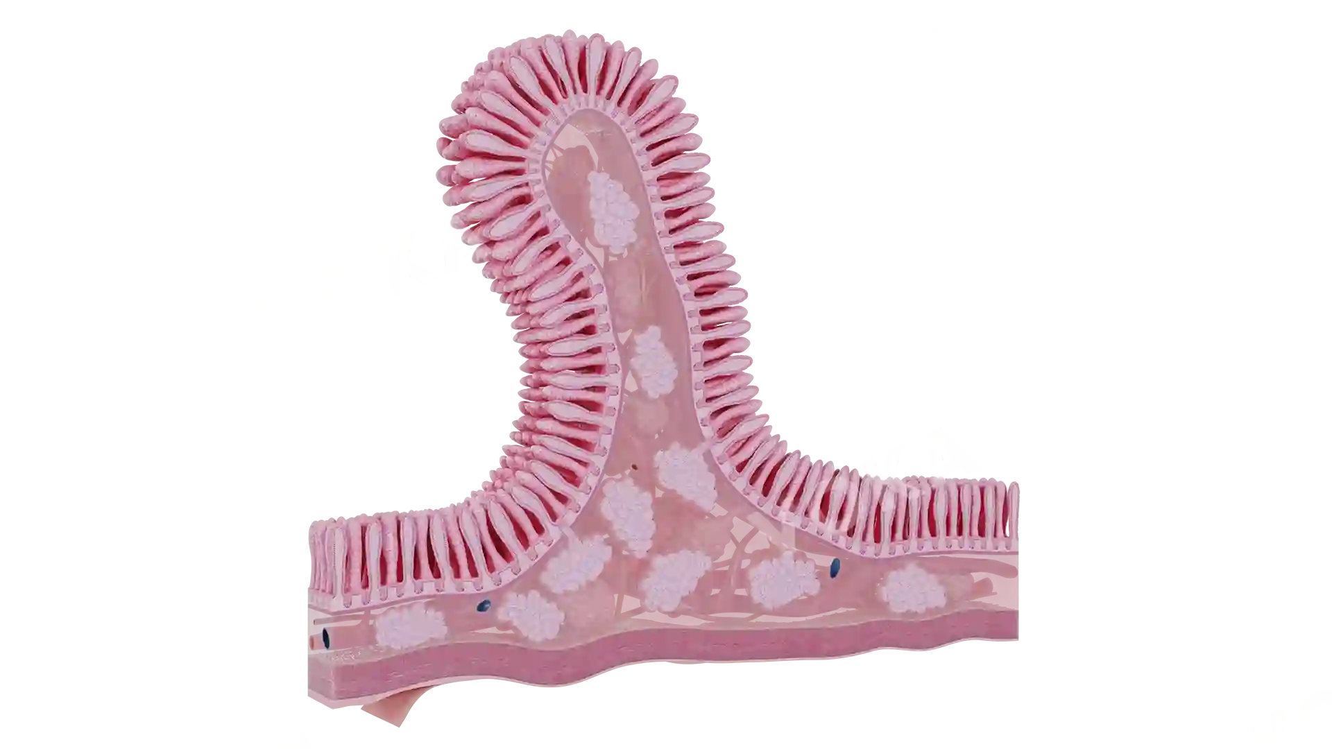
This anatomical image presents a clear view of a small duodenal ulcer located on the inner lining of the duodenum. The lesion appears as a well-defined, round, pale crater surrounded by slightly inflamed mucosa. The smooth folds of the intestinal wall are visible in contrast to the disrupted area, emphasizing the localized nature of the ulcer. The open sectional perspective provides a focused and educational depiction of early-stage peptic ulcer disease in the duodenum.
Related items


Small Duodenal Ulcer image - 30079
Gastroenterology
Select license
More information
Details
Background
Transparent
Resolution
1920 x 1080 px
Orientation
Horizontal
Format
PNG
File size
1.7 Mb
Upload date
June 10, 2025
Item successfully added to the cart