/Acute%20Otitis%20Media%20-%20Perforation%2C%202.webp)
/Acute%20Otitis%20Media%20-%20Perforation%2C%201.webp)


.webp)
Description
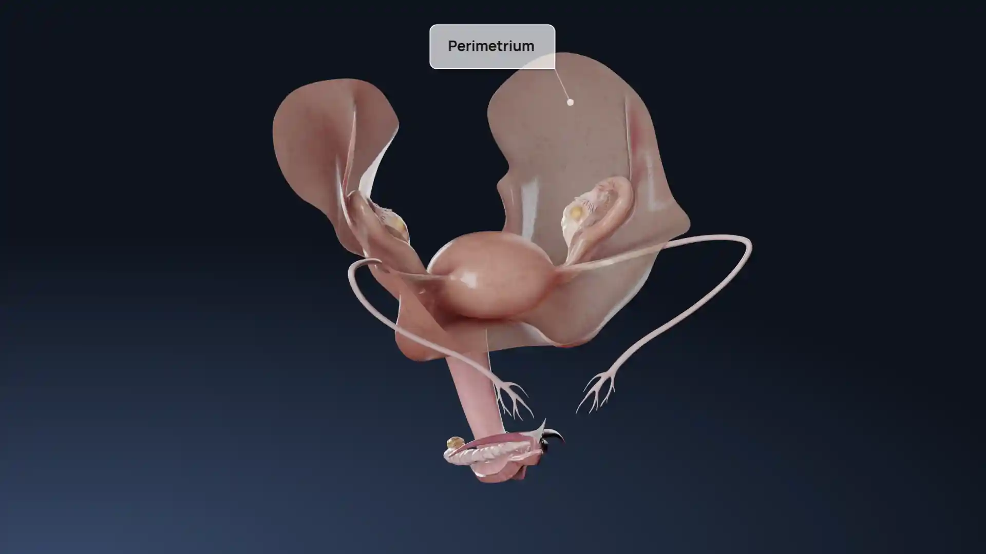
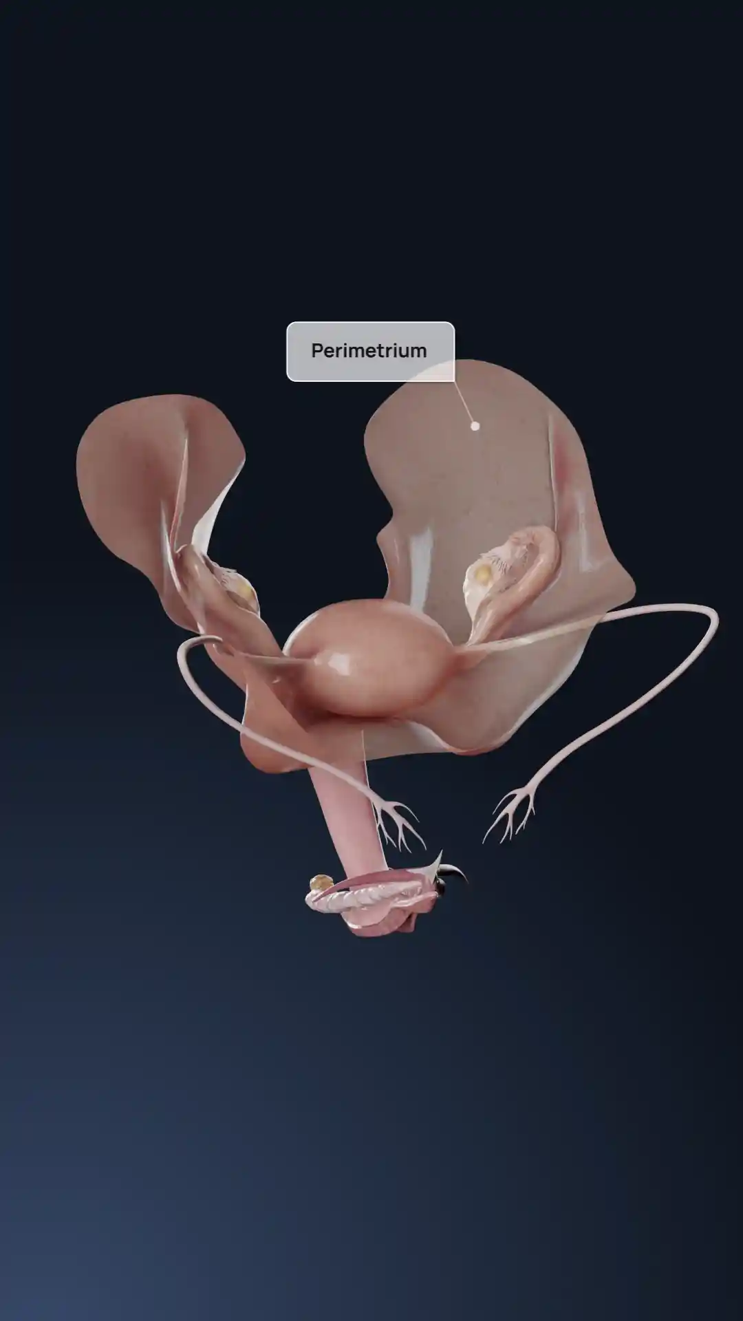
This anatomical illustration depicts a leiomyoma of the uterus, also known as a uterine fibroid, as classified according to the FIGO system. The image provides a cutaway view of the uterus and surrounding reproductive anatomy, including the ovaries and fallopian tubes, offering a clear context for the tumor’s position and relationship to the endometrium and serosa. The fibroid itself is shown as a distinct, rounded, pale mass that disrupts the smooth contour of the uterine wall. This type of visualization is beneficial for understanding the spatial anatomy of fibroids and their potential impact on menstruation, fertility, or adjacent organs, making it a valuable resource for medical education and gynecological consultations.
Related items


Leiomyoma of the uterus (FIGO Type) image - 30087
Gynecology
Select license
More information
Details
Item successfully added to the cart