/Acute%20Otitis%20Media%20-%20Perforation%2C%202.webp)
/Acute%20Otitis%20Media%20-%20Perforation%2C%201.webp)


Description
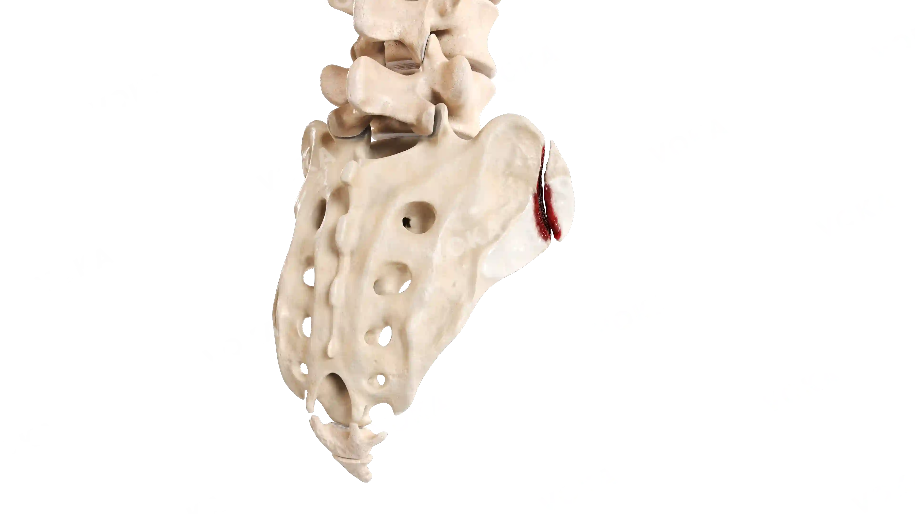
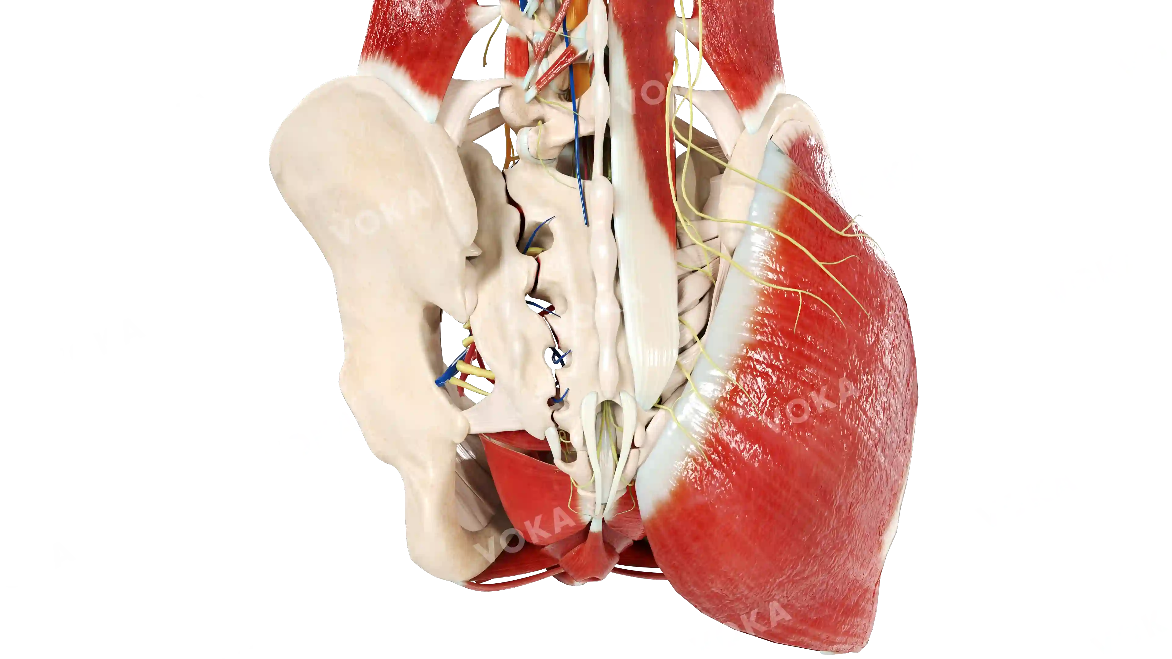
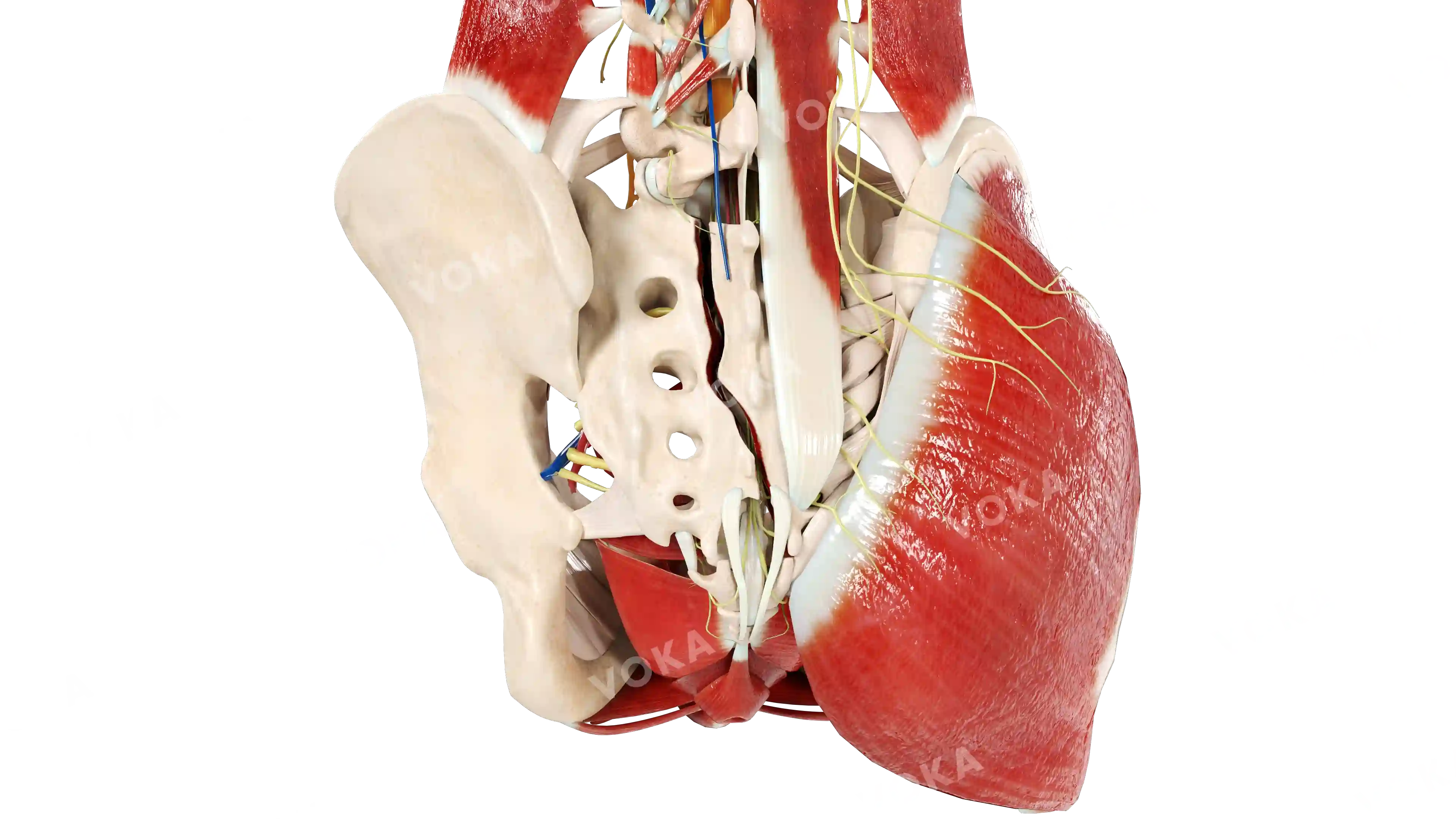
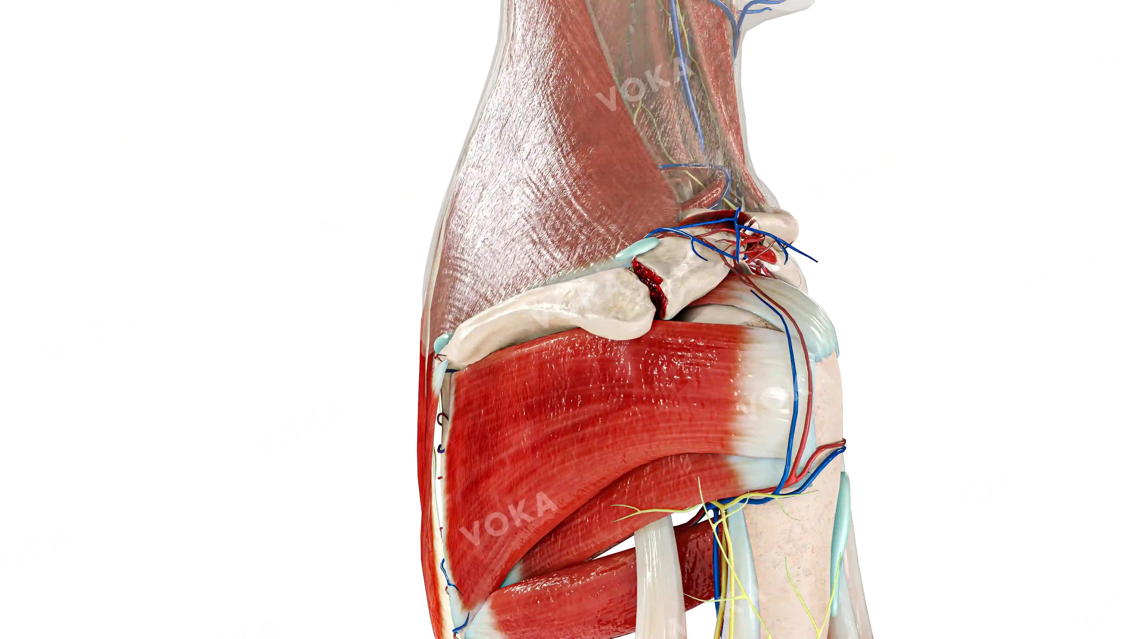
This high-resolution 3D visualization provides a detailed look at the atlas, the first cervical vertebra, offering a clear and comprehensive view of its unique structure. The image highlights key anatomical features, including the anterior and posterior arches, lateral masses, and the superior articular facets that articulate with the skull. Ideal for medical students, chiropractors, and educators, this resource is essential for presentations, classroom instruction, and clinical training. Its precise and engaging depiction simplifies complex spinal anatomy, making it easier to understand the atlas’s role in head movement and cervical stability.
Related items


Atlas, first cervical vertebra image - 25243
Musculoskeletal system
Select license
More information
Details
Item successfully added to the cart