/Acute%20Otitis%20Media%20-%20Perforation%2C%202.webp)
/Acute%20Otitis%20Media%20-%20Perforation%2C%201.webp)


Description
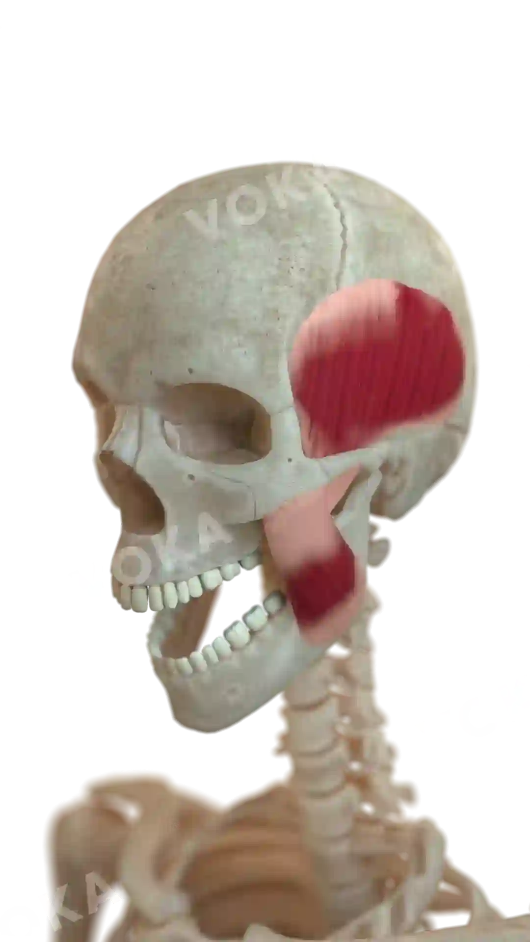
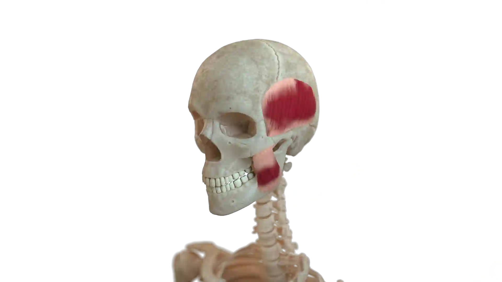
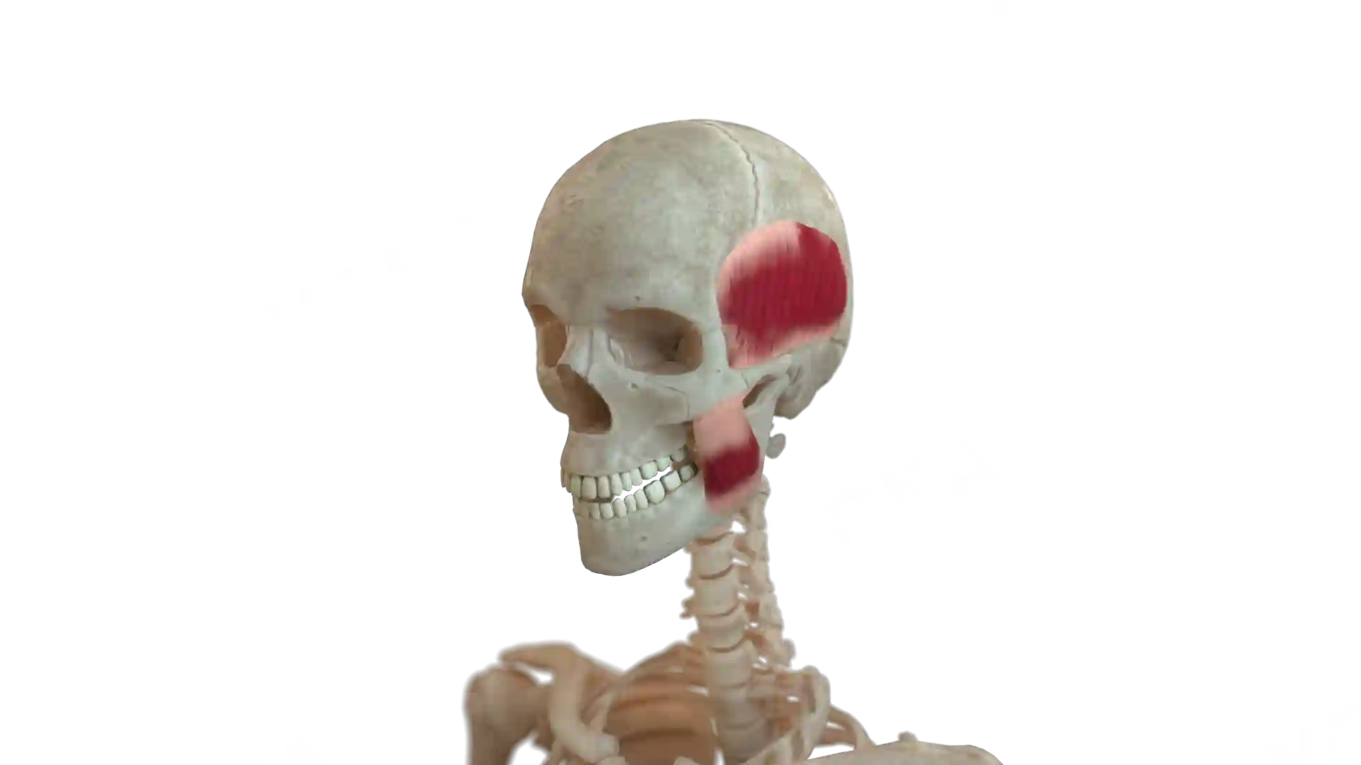
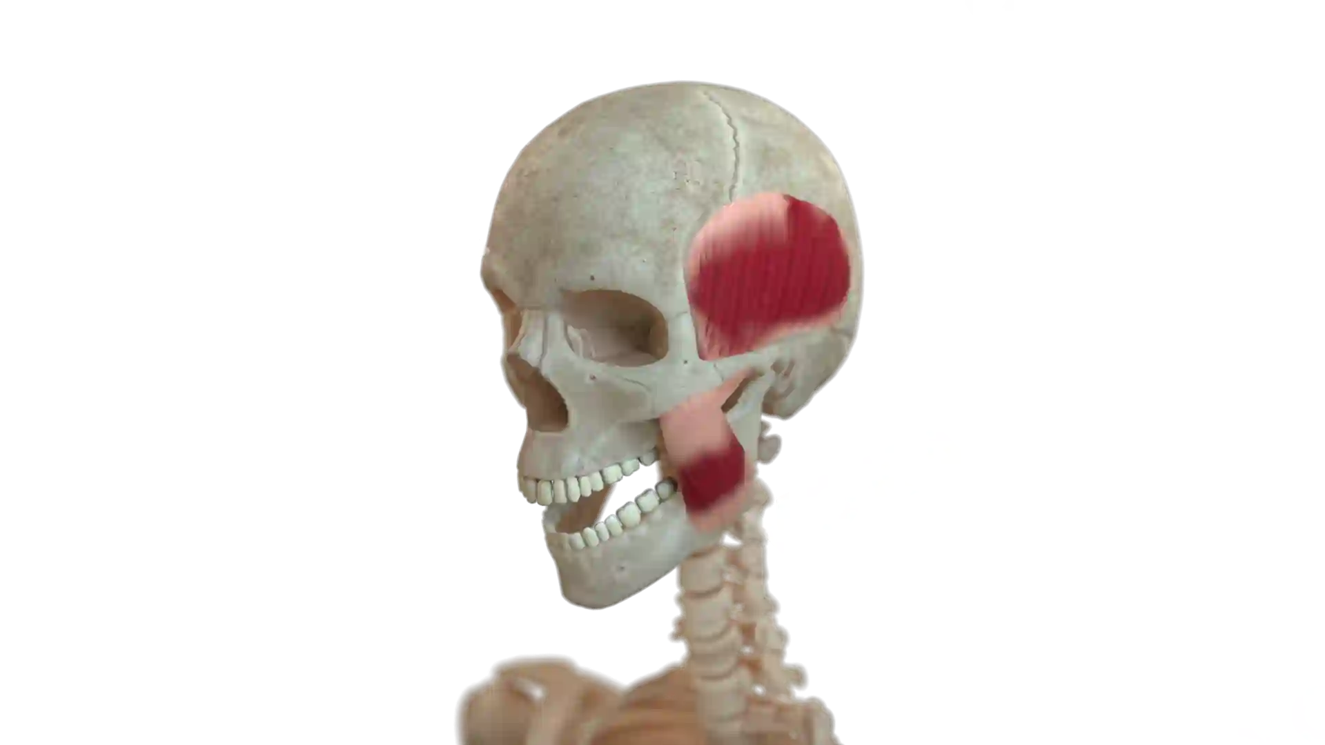
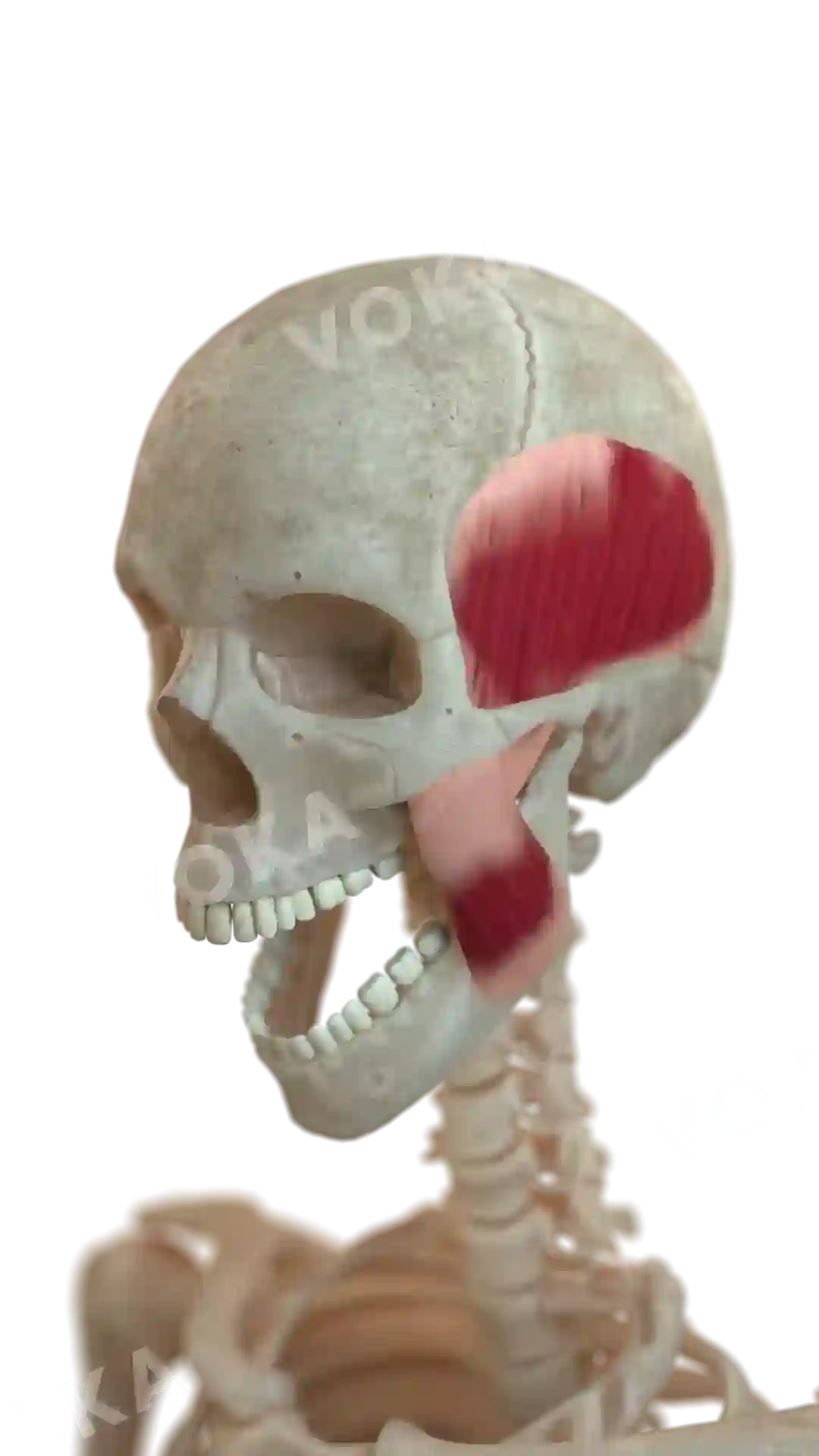
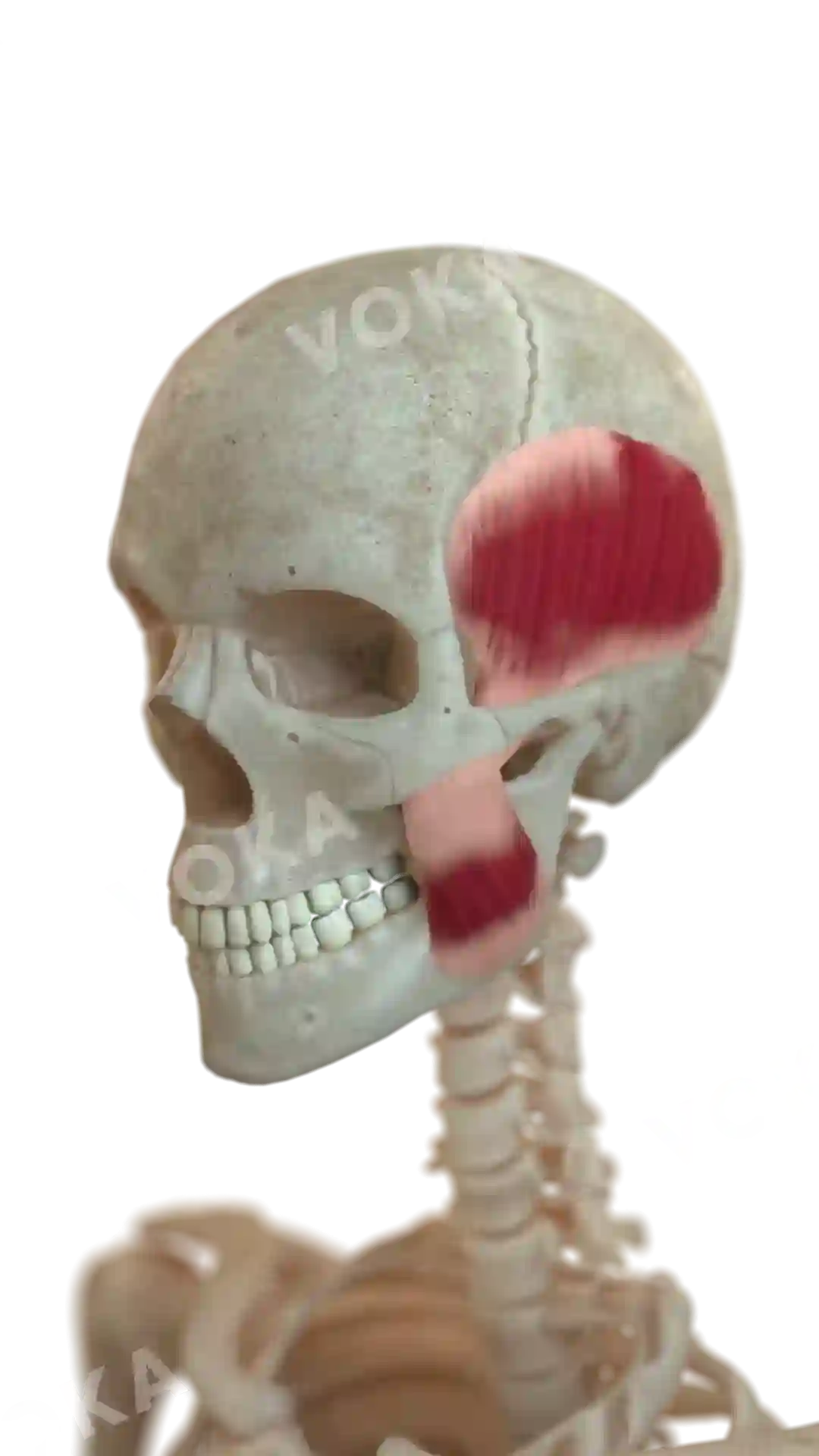
In this anatomical illustration, the human skull is presented with partially opened jaws, emphasizing the temporalis and masseter muscles. The temporalis muscle, fanned across the temple region, and the thick, rectangular masseter muscle are vividly colored to showcase their significance in jaw movement. The image provides a dynamic perspective, highlighting the interaction between bone and muscle during mastication. This visual aid is perfect for medical students, educators, and health professionals, offering an insightful look into the biomechanics of chewing, muscle attachment points, and functional anatomy.
Related items

Head muscles image - 25281
Musculoskeletal system
Select license
More information
Details
Background
Transparent
Resolution
1080 x 1920 px
Orientation
Vetical
Format
PNG
File size
1.4 Mb
Upload date
February 27, 2025
Item successfully added to the cart