/Acute%20Otitis%20Media%20-%20Perforation%2C%202.webp)
/Acute%20Otitis%20Media%20-%20Perforation%2C%201.webp)


Description
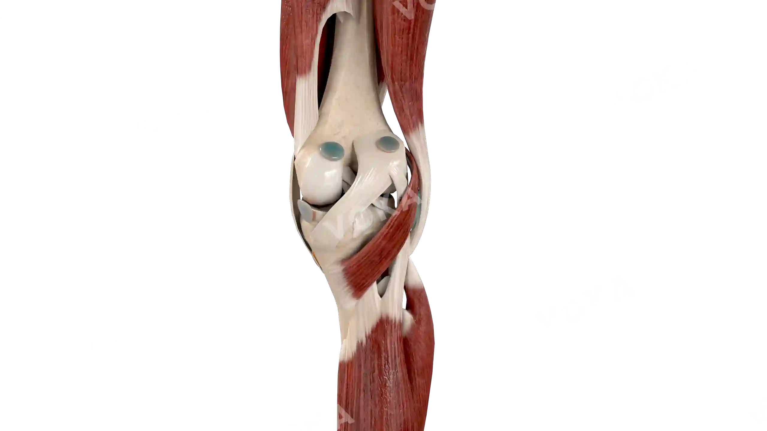
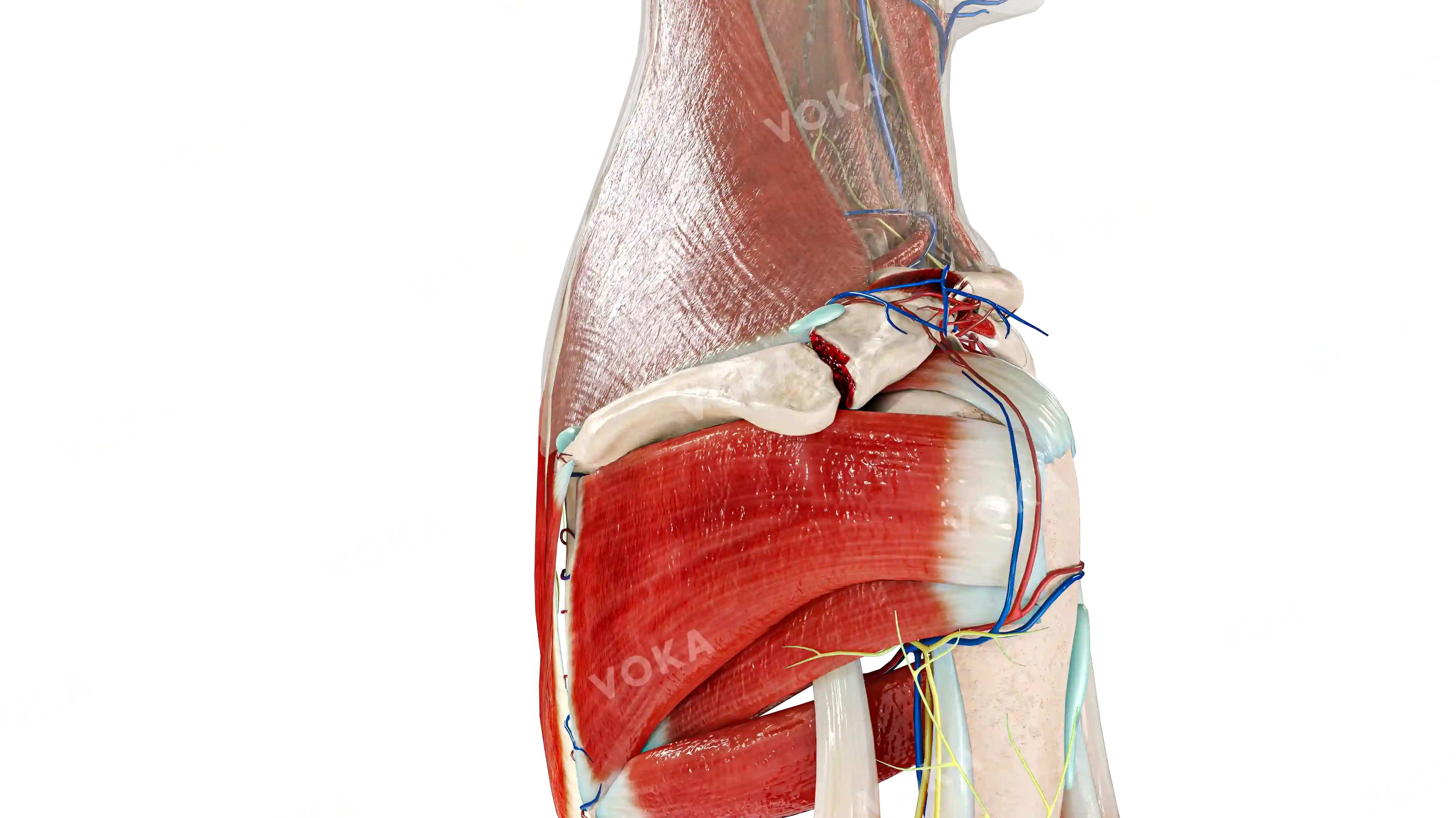
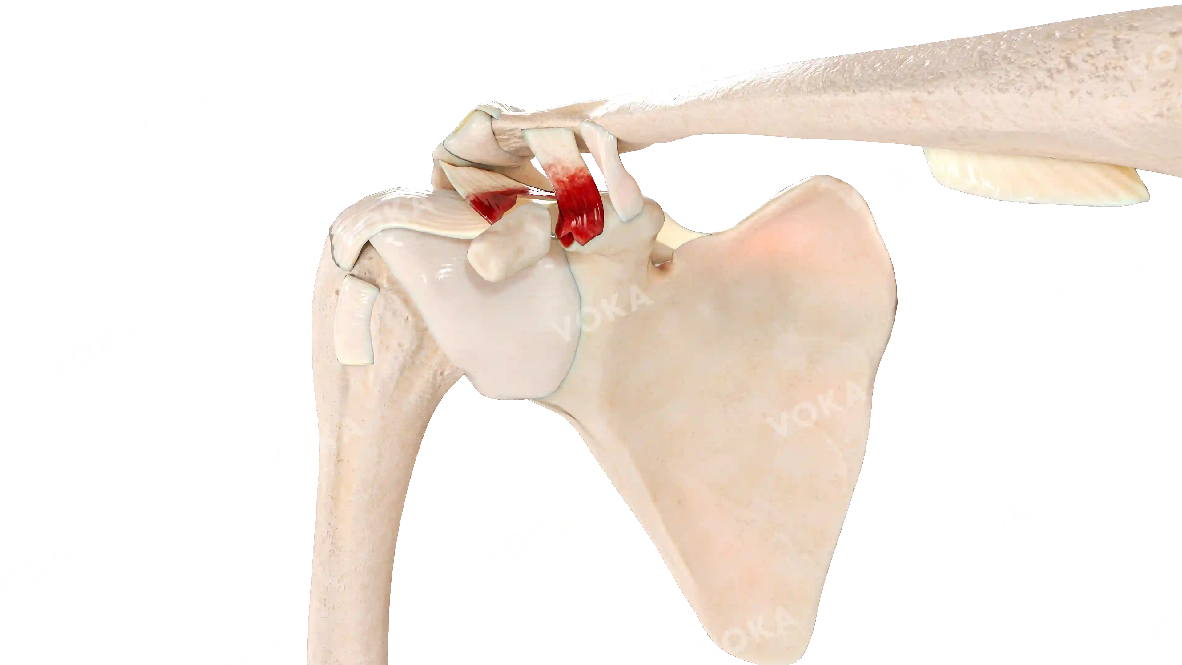
This detailed anatomical illustration showcases the complex structure of the knee joint, emphasizing the integration of muscles, ligaments, bones, and cartilage. The prominent femur, tibia, and patella are clearly visible, with surrounding ligaments like the cruciate ligament and the patellar tendon providing stability. The illustration highlights the quadriceps muscle group, partially covering the joint and playing a vital role in knee extension. This image serves as an invaluable resource for understanding knee biomechanics, ideal for medical education and clinical reference.


Knee with muscles, right image - 25301
Musculoskeletal system
Select license
More information
Details
Background
Transparent
Resolution
2560 x 1440 px
Orientation
Horizontal
Format
PNG
File size
1.5 Mb
Upload date
February 27, 2025
Item successfully added to the cart