/Acute%20Otitis%20Media%20-%20Perforation%2C%202.webp)
/Acute%20Otitis%20Media%20-%20Perforation%2C%201.webp)


Description
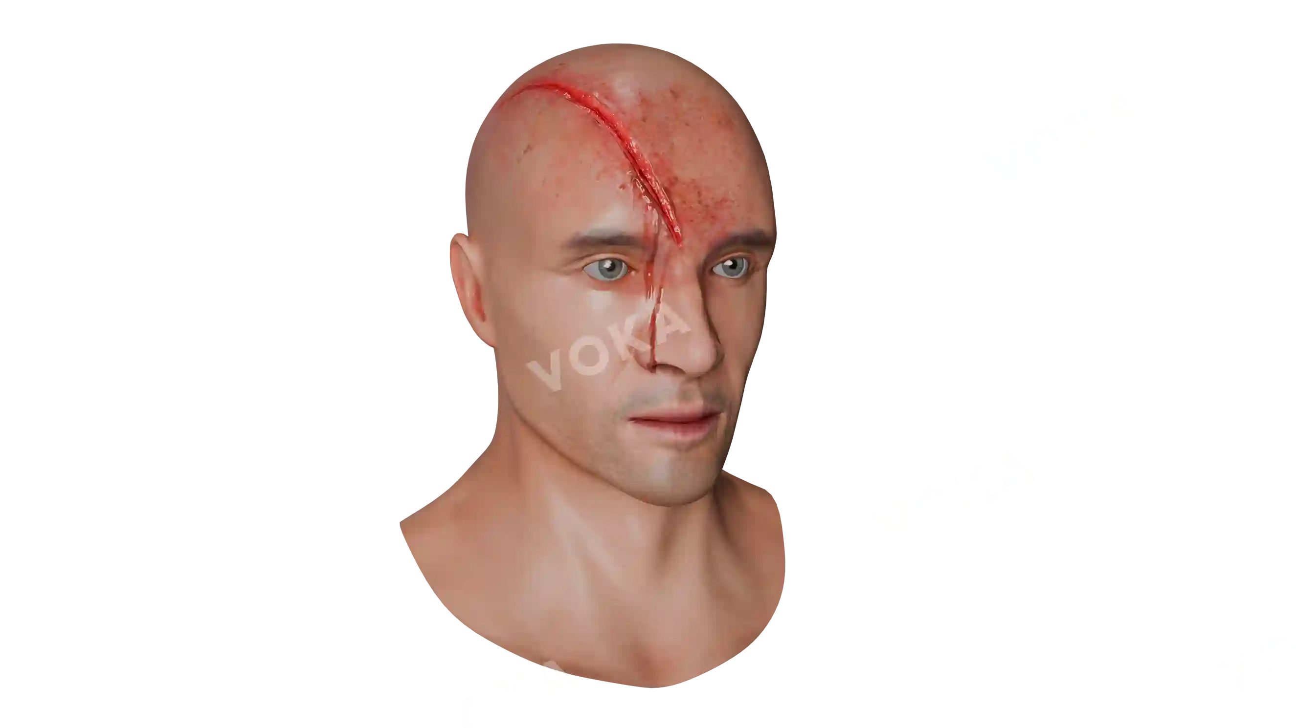
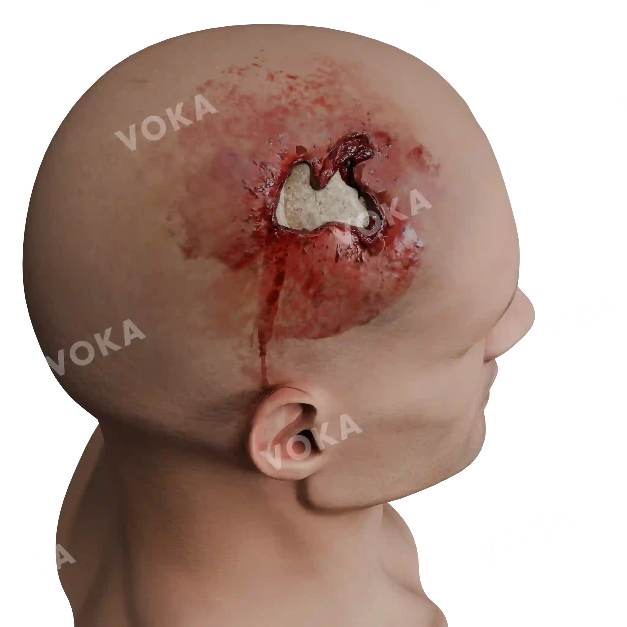
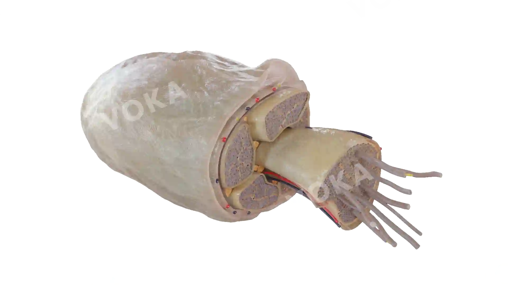
This vivid 3D anatomical model illustrates a subdural hemorrhage, an accumulation of blood between the dura mater and the brain’s surface. The image shows a substantial pooling of dark red blood compressing the cerebral hemisphere, simulating the mass effect and midline shift commonly associated with severe cases. Fractured cranial bones and disrupted vasculature emphasize the traumatic origin of the injury. Ideal for neurology education, trauma simulation, and forensic pathology training, this model highlights the clinical urgency of subdural hematomas and their potential impact on brain function and patient outcomes.
Related items


Subdural hemorrhage image - 30125
Neurology
Select license
More information
Details
Background
Transparent
Resolution
1920 x 1080 px
Orientation
Horizontal
Format
PNG
File size
8.3 Mb
Upload date
June 10, 2025
Item successfully added to the cart