/Acute%20Otitis%20Media%20-%20Perforation%2C%202.webp)
/Acute%20Otitis%20Media%20-%20Perforation%2C%201.webp)


.webp)
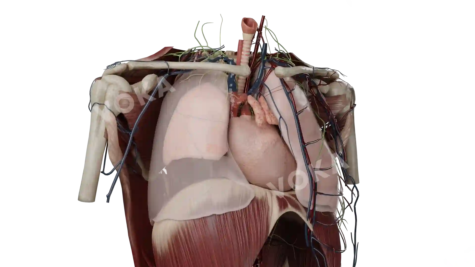
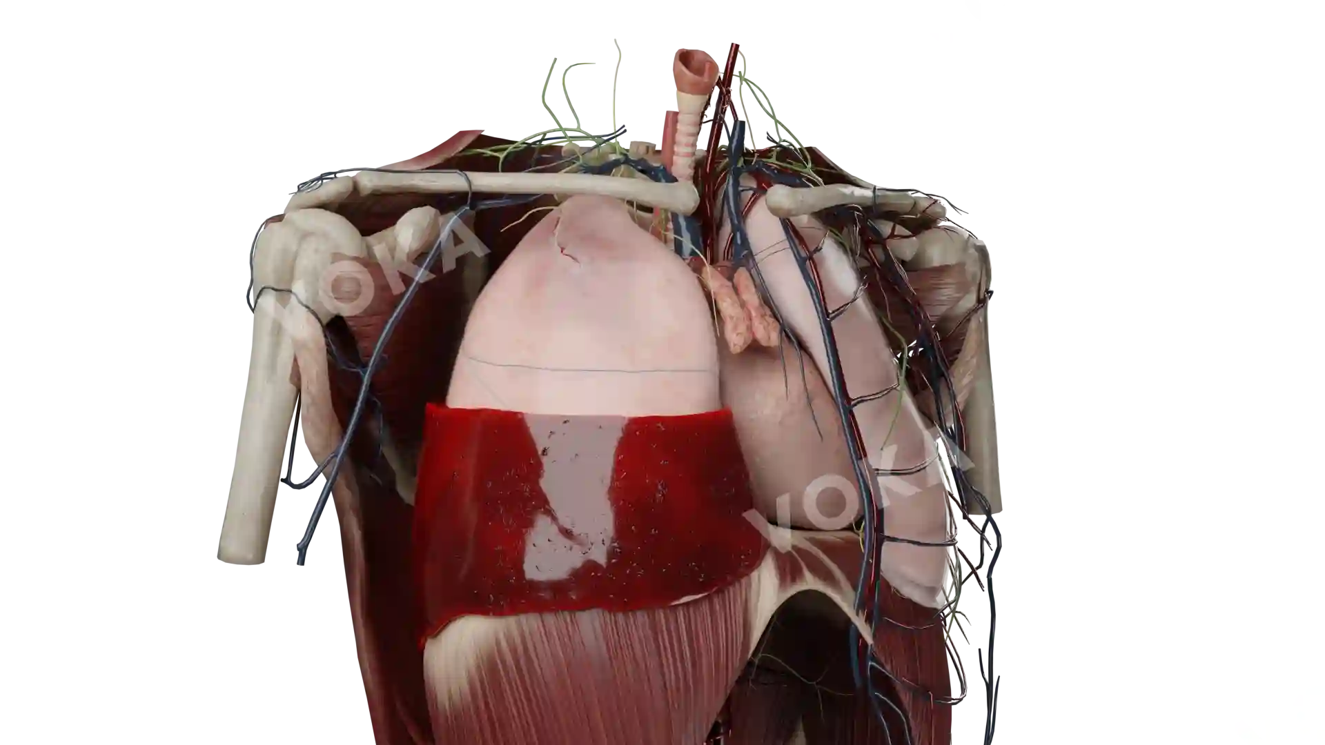
Description
This close-up anatomical illustration provides a detailed view of the tracheal wall, highlighting its layered structure. The image emphasizes the cartilage rings, smooth muscle, and mucosal lining that form the trachea's protective barrier and aid in air conduction. The depiction reveals how the tracheal wall maintains the rigidity required for airflow while remaining flexible enough for respiratory movements. Ideal for respiratory system studies, this image offers valuable insights for medical professionals, students, and researchers focused on respiratory health, diseases, and treatments.
Related items


.webp)


Tracheal wall (close up) image - 25226
Pulmonology
Select license
More information
Details
Background
Transparent
Resolution
1920 x 1080 px
Orientation
Horizontal
Format
PNG
File size
15.8 Mb
Upload date
February 27, 2025
Item successfully added to the cart