/Acute%20Otitis%20Media%20-%20Perforation%2C%202.webp)
/Acute%20Otitis%20Media%20-%20Perforation%2C%201.webp)


September 11, 2025
Description
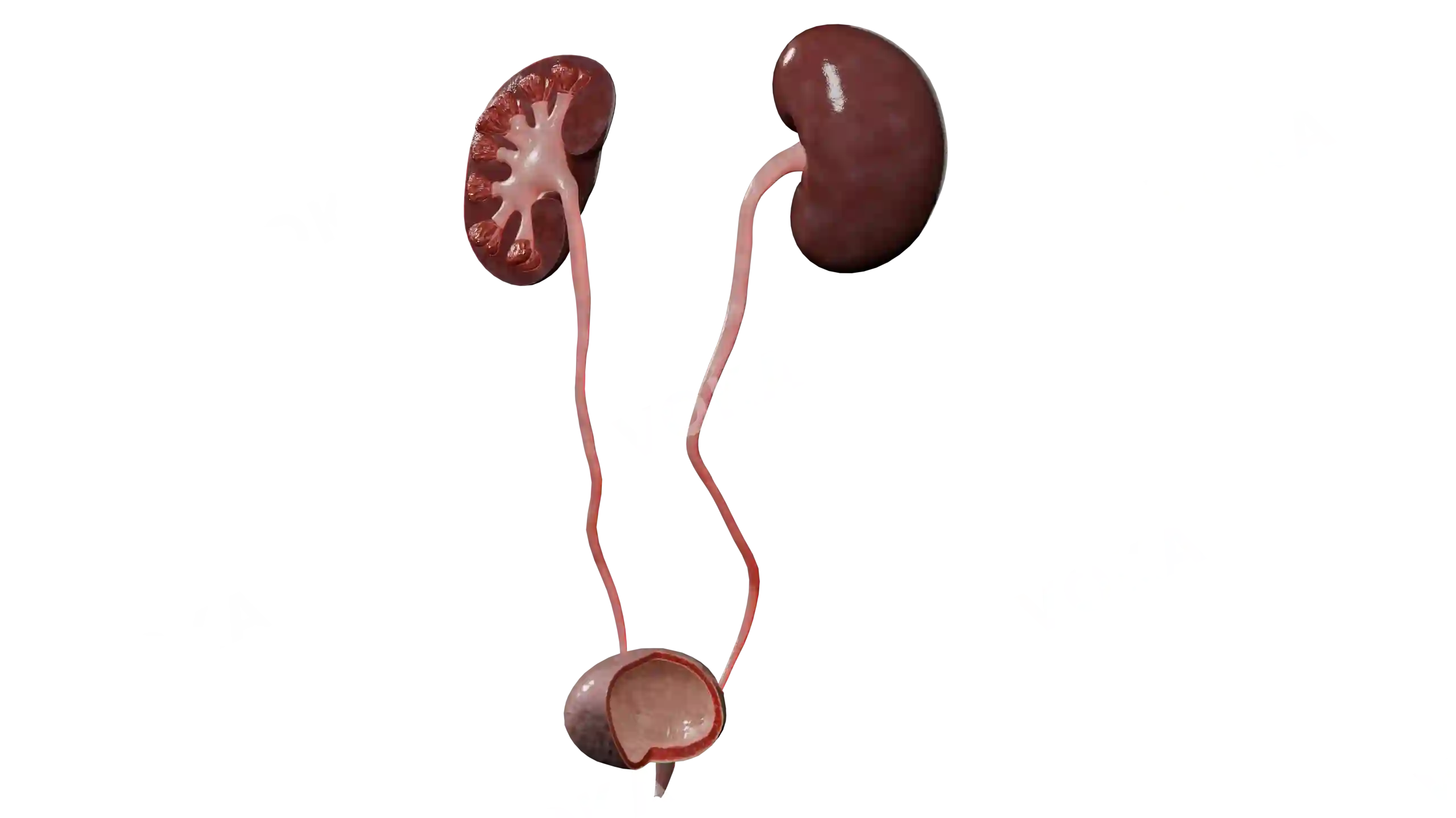
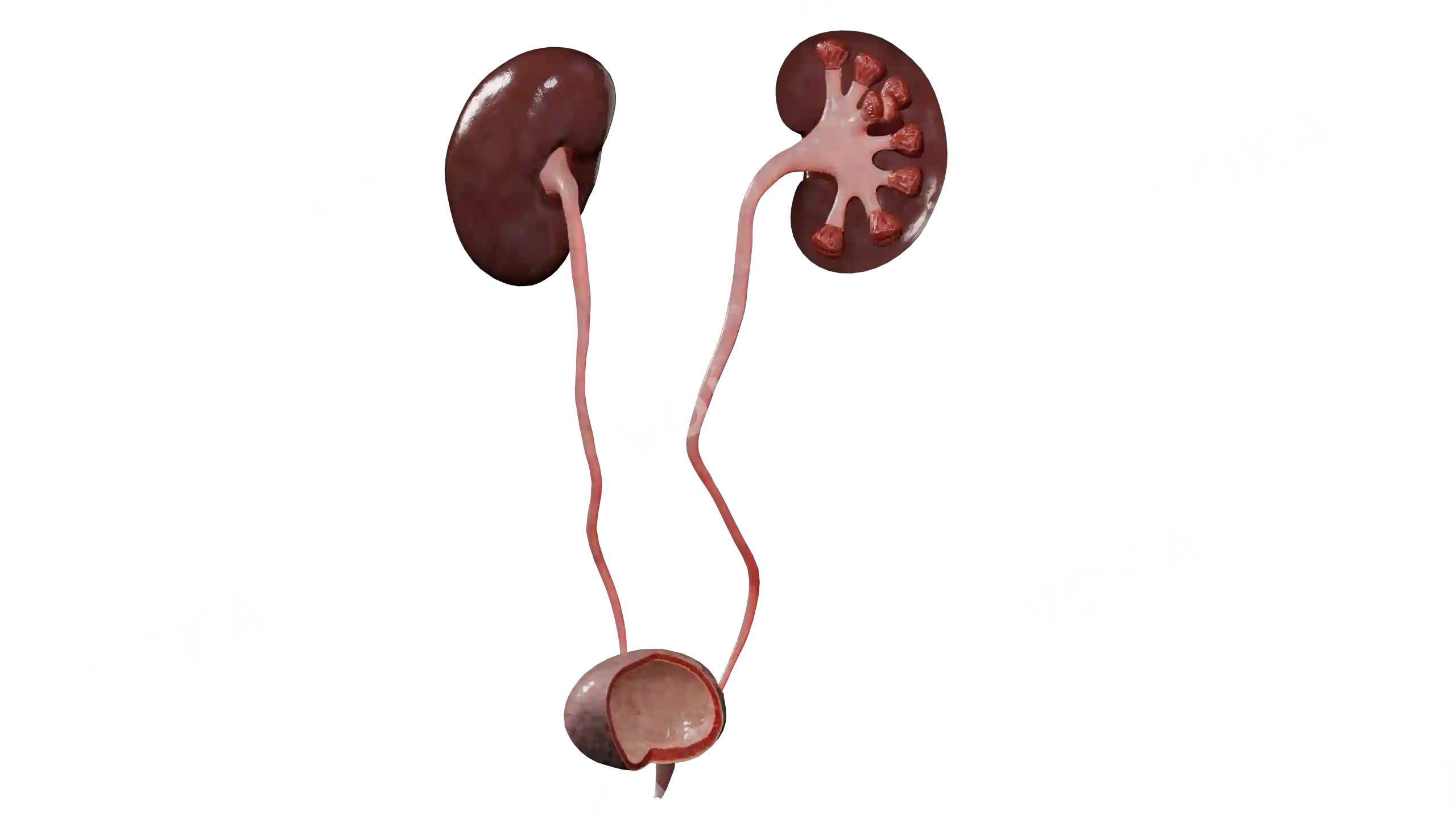
Visualize the internal anatomy of the right kidney and associated urinary tract structures in this 3D frontal section model. It displays the renal cortex, medulla, pyramids, calyces, pelvis, and ureter in their correct spatial relationships. With this model, learners can trace how urine is collected from nephrons and funneled into the ureter. This anatomical view helps students understand the organization of the urinary system and provides a foundation for exploring pathological changes.
Related items
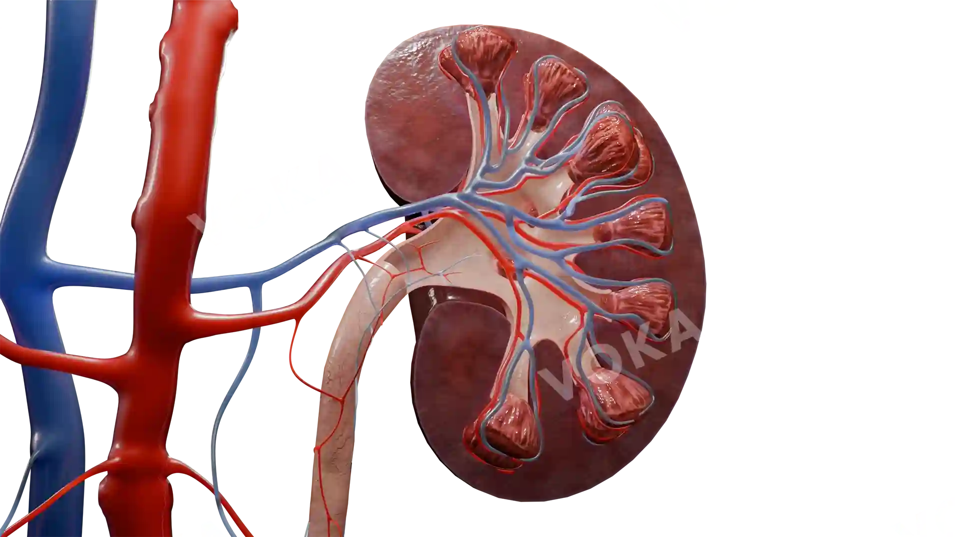
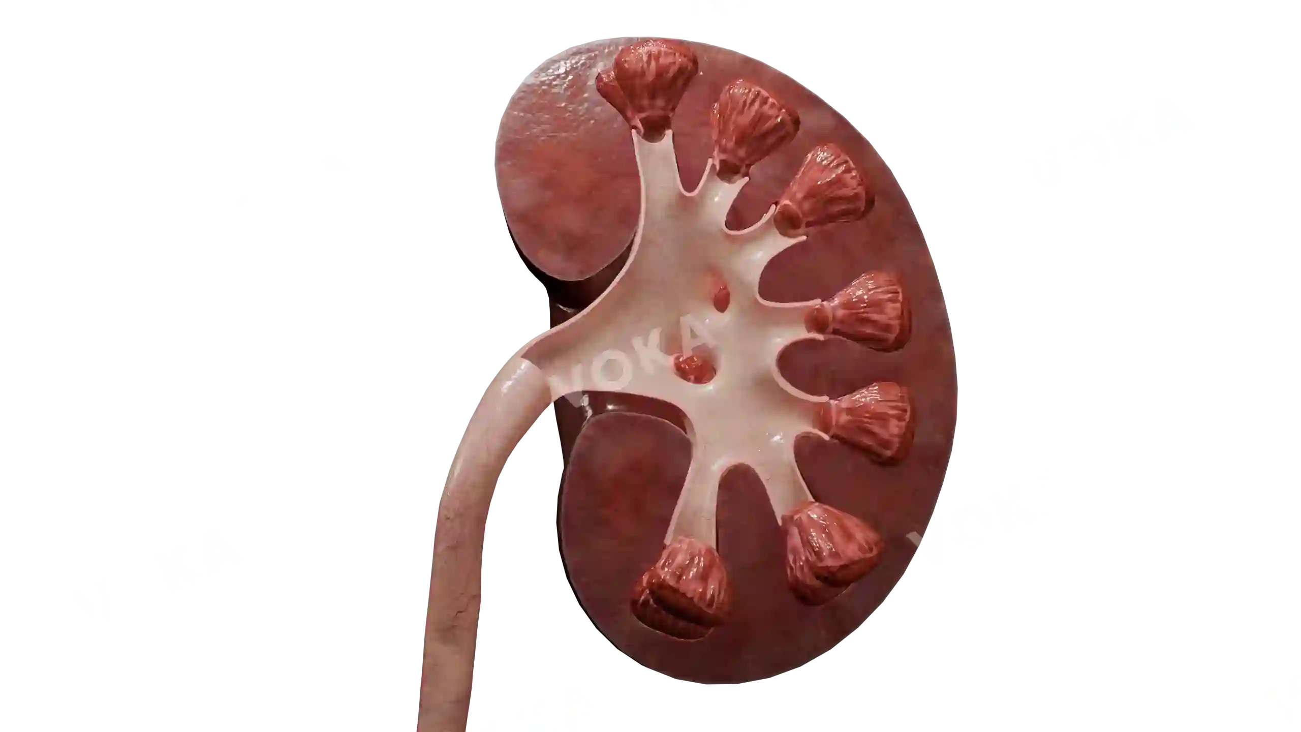

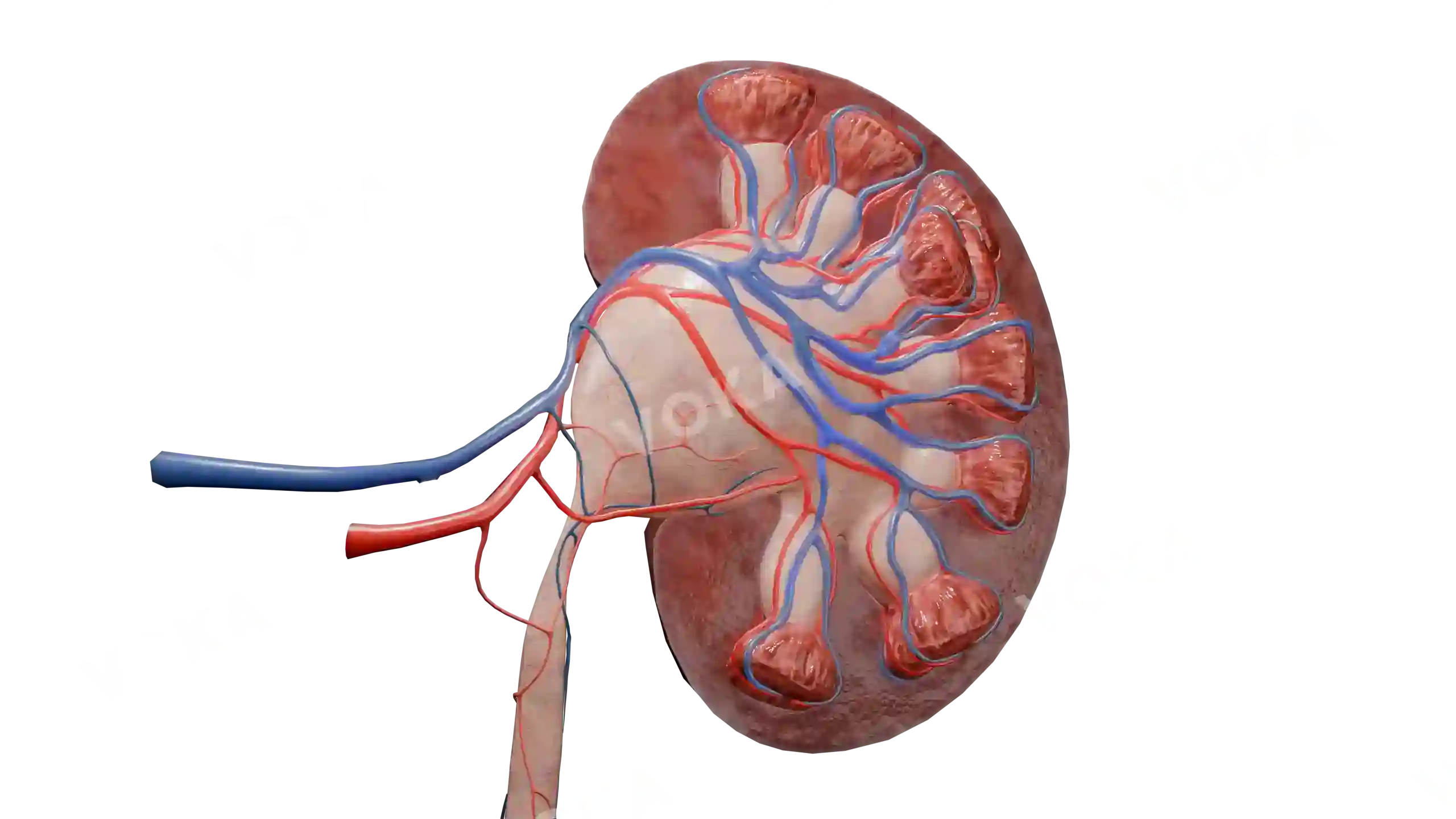


Urinary tract: right kidney in frontal section image - 30474
Urology
September 17, 2025
Select license
More information
Details
Background
Transparent
Resolution
2560 x 1440 px
Orientation
Horizontal
Format
PNG
File size
14.8 Mb
Item successfully added to the cart