/Acute%20Otitis%20Media%20-%20Perforation%2C%202.webp)
/Acute%20Otitis%20Media%20-%20Perforation%2C%201.webp)


Description
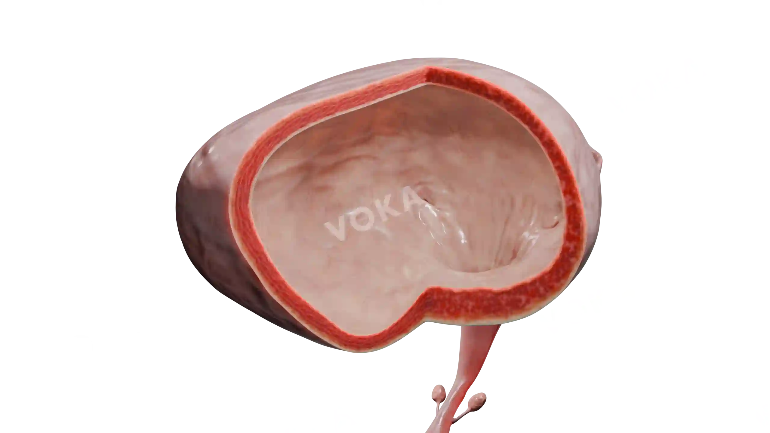
Check out the structure of a renal angiomyolipoma with this detailed 3D model. It shows the benign tumor composed of blood vessels, smooth muscle, and fat, which often develops within the renal cortex. The model highlights the heterogeneous composition of the lesion and its spatial relation to surrounding renal tissue. By studying this visualization, learners can understand how such growths may remain asymptomatic or lead to complications such as bleeding or mass effect.
Related items
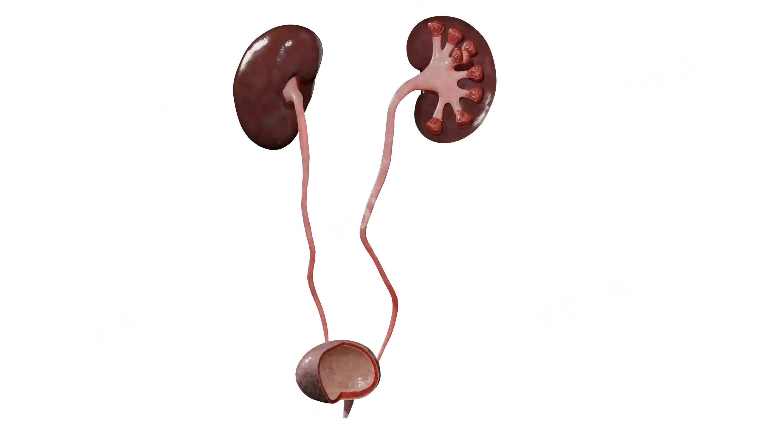
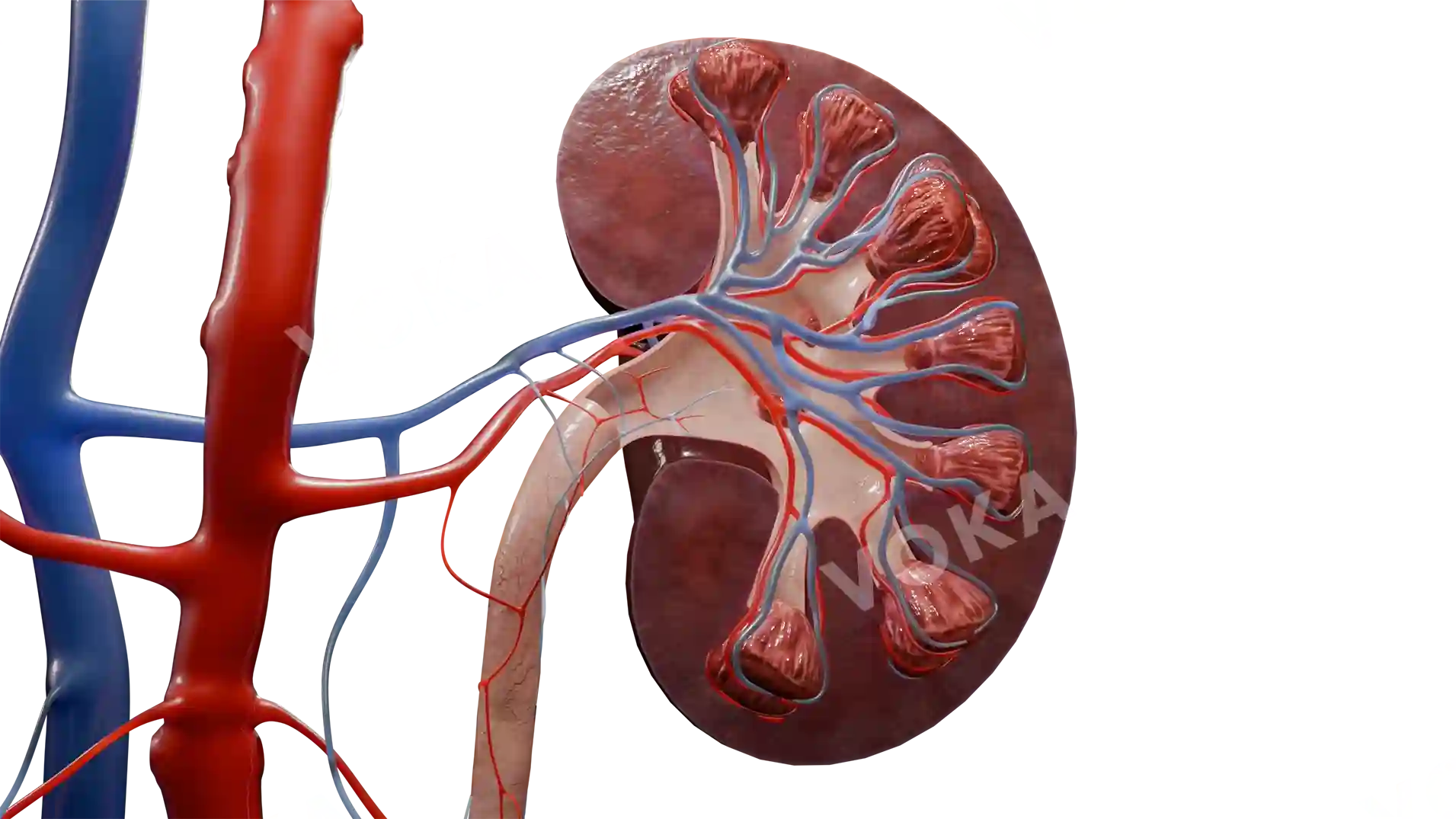
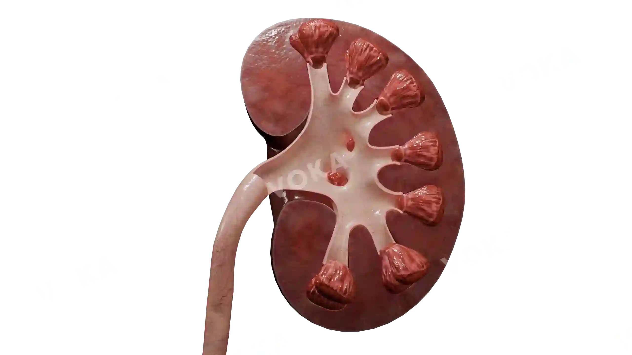


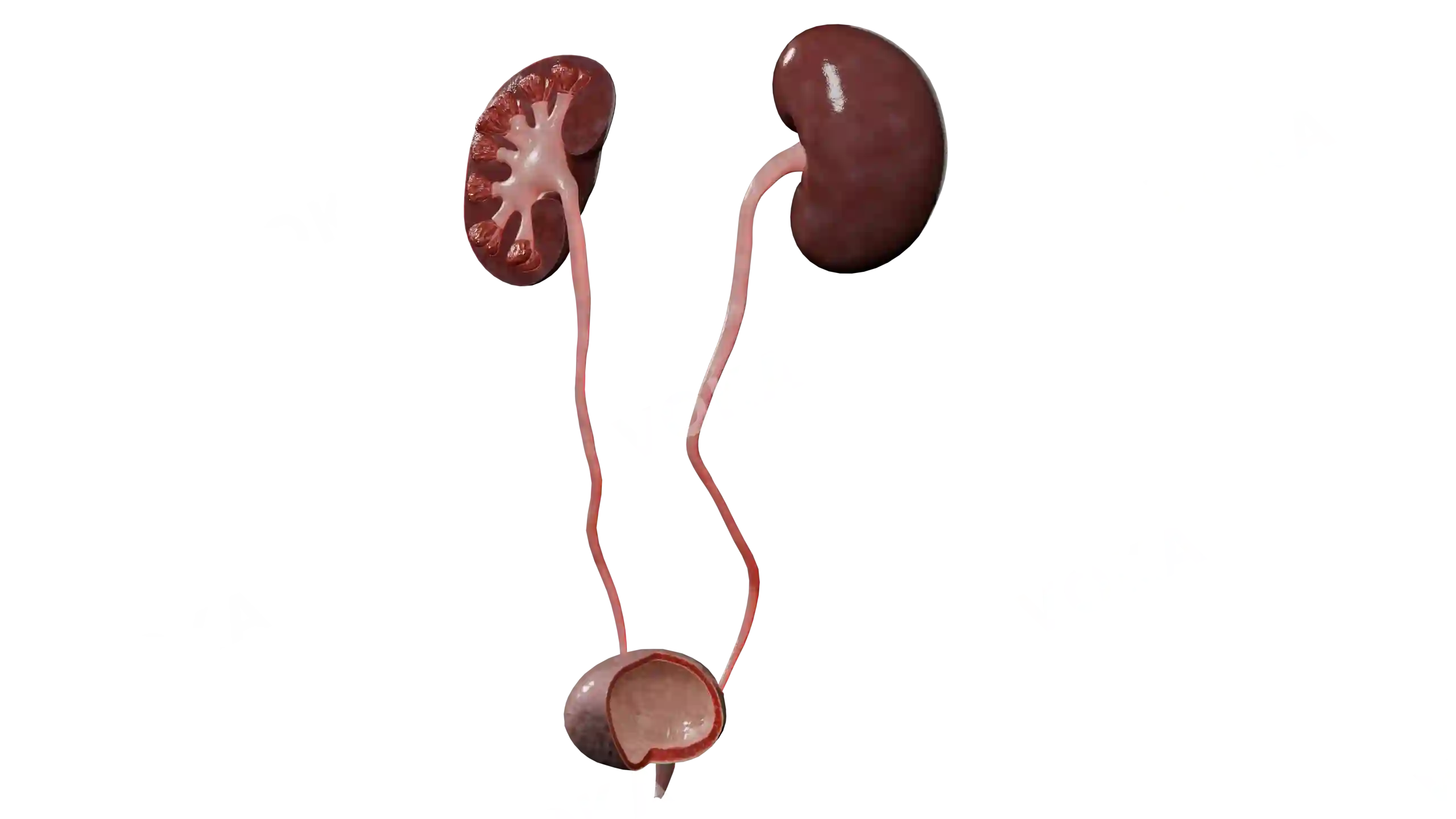
Renal angiomyolipoma image - 30472
Urology
Select license
More information
Details
Background
Transparent
Resolution
1920 x 1080 px
Orientation
Horizontal
Format
PNG
File size
8.3 Mb
Upload date
September 17, 2025
Item successfully added to the cart